 Log In
Search




 Open Access
 linmao@usst.edu.cn
Lin Mao, Shanghai Institute for Minimally Invasive Therapy, School of Health Science and Engineering, University of Shanghai for Science and Technology, No. 516 Jungong Road, Yangpu District, Shanghai 200093, China. Tel: +86-21-55572159. E-mail: linmao@usst.edu.cn.
Received November 28, 2025; Accepted February 7, 2026; Published June 17, 2026
● Current innovation in gastrointestinal defect management falls into two main categories: clip-based mechanical compression devices and advanced high-precision endoscopic suturing systems.
● Through-the-scope clips combined with auxiliary devices offer a strategic solution to overcome the size limitations inherent to single-device closure strategies for larger or complex defects.
● Future technological development should focus on enhancing reliability, operability, cost-effectiveness, and overall user-friendliness of advanced endoscopic closure devices to broaden their clinical applicability.
Open Accesslinmao@usst.edu.cnLin Mao, Shanghai Institute for Minimally Invasive Therapy, School of Health Science and Engineering, University of Shanghai for Science and Technology, No. 516 Jungong Road, Yangpu District, Shanghai 200093, China. Tel: +86-21-55572159. E-mail: linmao@usst.edu.cn.
Received November 28, 2025; Accepted February 7, 2026; Published June 17, 2026
● Current innovation in gastrointestinal defect management falls into two main categories: clip-based mechanical compression devices and advanced high-precision endoscopic suturing systems.
● Through-the-scope clips combined with auxiliary devices offer a strategic solution to overcome the size limitations inherent to single-device closure strategies for larger or complex defects.
● Future technological development should focus on enhancing reliability, operability, cost-effectiveness, and overall user-friendliness of advanced endoscopic closure devices to broaden their clinical applicability.
Advanced endoscopic therapeutic techniques, such as endoscopic mucosal resection, endoscopic submucosal dissection, and third-space endoscopy, have transformed digestive endoscopy into a minimally invasive surgical platform. This transformation, nonetheless, poses a major challenge, namely the safe and efficient closure of the postoperative gastrointestinal (GI) defects that arise, as inadequately closed defects may lead to serious postoperative complications. This review provides a comprehensive overview of the historical background, the basic principles of closure, and the technical changes of the mainstream devices used for closure of GI defects, viewed mostly from the perspective of medical devices and engineering design. We have divided the most significant closure devices currently in use into endoscopic clipping devices and endoscopic suturing devices, and we have outlined their respective historical development and working principles. In addition, this review has identified and explained advanced clip-based closure methods that employ multiple through-the-scope clips (TTSCs) or are combined with auxiliary devices to overcome the inherent limitations of single-TTSC use. This review aims to provide endoscopists with a deeper understanding of the existing closure devices so as to facilitate their optimal clinical use, and to offer insights that may help in the development of next-generation closure technologies.
Keywords: Endoscopy, Postoperative defects, Through-the-scope clip, Over-the-scope clip, Endoscopic suturing device
Digestive endoscopy has evolved beyond tissue sampling and excision to become a surgical platform for delivering therapeutic devices to treat a variety of gastrointestinal (GI) disorders [1]. The emergence of techniques like endoscopic mucosal resection (EMR) and endoscopic submucosal dissection (ESD) has made the resection of early GI tumors possible [2, 3]. The popularization of endoscopic full-thickness resection (EFTR) and of third-space endoscopy techniques such as peroral endoscopic myotomy, have established digestive endoscopy as a natural orifice transluminal endoscopic surgery platform [4-6].
While these advanced techniques offer immense clinical benefits, a significant challenge arises: the management of complicated postprocedural defects [7-9]. Endoscopic resection procedures inevitably create large, deep, or even full-thickness defects [10, 11]. Postoperative defects, especially iatrogenic endoscopic perforations, can lead to extremely severe complications [11, 12]. Therefore, the timely closure of defects can effectively reduce postoperative adverse events [13, 14]. Traditional closure devices are often insufficient for complex defects. To meet these challenges, endoscopic closure devices and related technologies have evolved in parallel with advancements in endoscopic surgery [15]. The endoscopist’s armamentarium has expanded from the early through-the-scope clips (TTSCs) for closing small defects, to over-the-scope clips (OTSCs) with greater closure strength, and subsequently to endoscopic suturing devices capable of achieving “surgical-style” closure.
To date, many reviews have introduced commercially available closure devices from a clinical perspective, providing strategies for device selection based on different types of GI defects [16-20]. This review, however, focuses on the development process and working principles of closure devices. It reviews the closure principles and development history of endoscopic clipping devices and endoscopic suturing devices, and also introduces advanced clip-based closure methods that combine TTSCs with auxiliary devices. This review aims not only to guide endoscopists in the rational selection and utilization of their armamentarium but also to provide insights for both clinicians and engineers to develop next-generation closure devices and methods based on current instrumentation.
2.1 TTSCs
Since their first report by Hayashi et al. in 1975, TTSCs have evolved for approximately 50 years [21]. As indications have continuously expanded, TTSCs are now the most common and widely used closure devices in diagnostic and therapeutic digestive endoscopy (Figure 1) [22]. A regular TTSC comprises three key components: a metallic clip for tissue approximation, a delivery catheter based on tendon-sheath transmission, and an operating handle to control clip deployment [23, 24]. All TTSCs are passed through the instrument channel and exit at the distal end of the endoscope to reach the target lesion.


Figure 1. Different types of through-the-scope endoscopic clips. (A) Resolution 360 (Boston Scientific); (B) Instinct Plus (Cook Medical); (C) EZ Clip (Olympus); (D) SureClip (Micro-Tech); (E) DuraClip (ConMed). This figure is cited from [136].
The basic working principle of TTSCs is to compress tissue by using the mechanical force generated upon closure, thereby achieving hemostasis or the approximation of defect edges. Different brands and models of TTSCs possess specific geometrical parameters, physical properties, or additional features, such as rotatability, compression strength, and compatibility with magnetic resonance imaging (Table 1) [25, 26].

Early applications of TTSCs demonstrated favorable therapeutic results, but a series of limitations hindered their clinical adoption, such as cumbersome manipulation, low clip-retention rate, and the inability to rotate the clip to the designated axial direction [27-29]. In the 1990s, Hachisu et al., in collaboration with Olympus (Tokyo, Japan), developed a prototype and a clinical model of the rotatable TTSC, which improved the device’s maneuverability [30]. In the 21st century, TTSC development has concentrated on integrating enhanced functional characteristics, including pre-loading, rotatability, re-opening capability, and reliable deployment [31]. Representative examples include the QuickClip series (Olympus, Tokyo, Japan), the Resolution Clip (Boston Scientific, Massachusetts, USA), the Instinct Clip (Cook Medical, NC, USA), and the SureClip (Micro-Tech, Nanjing, China) [25, 32, 33].
Most TTSCs consist of two prongs, whereas in 2003 Cook Medical (Winston-Salem, NC, USA) introduced the three-pronged TriClip, designed to more easily grasp protruding lesions and potentially reduce the number of clips used [34]. However, due to its higher cost and reportedly inferior hemostatic effect compared to regular TTSCs, two-pronged TTSCs remain the preferred choice [35].
Although the clip components themselves are typically disposable, TTSC systems can be categorized based on the reusability of the delivery system into single-use disposable systems and reusable systems with a reloadable applicator. Common reusable TTSCs include the EZ Clip (Olympus Medical Systems Corp., Tokyo, Japan) and the ZEOCLIP (Zeon Medical, Tokyo, Japan). The EZ Clip was one of the earliest reloadable, rotatable TTSCs, while the ZEOCLIP reportedly achieves a larger opening width faster and easier than the EZ Clip [36]. Furthermore, the ZEOCLIP can be modified into a fluorescent marking clip for tumor localization [37]. The recently reported re-openable SB clip (Sumitomo Bakelite Co., Ltd., Tokyo, Japan) also belongs to the category of reusable TTSCs [38]. Its deployment, rotation, and price are comparable to the EZ Clip, and it can close large mucosal defects via hold-and-drag closure [39].
The above-mentioned TTSC systems, whether disposable or reloadable, typically deploy only one clip before the catheter must be reloaded or replaced. In contrast, multi-firing TTSCs are less common, such as the InScope Multi-Clip Applier (Ethicon Endo-Surgery, Cincinnati, Ohio, USA) and the Clipmaster3 (Medwork, GmbH, Aisch, Germany) [40, 41]. These devices make it possible to insert multiple clips during one single operation, and thus a withdrawal and reload are not necessary for every clip [40, 41]. However, a related article reported that multi-firing TTSCs might not actually shorten the time of the procedure, probably due to the limited experience, and their effectiveness still needs to be confirmed by more clinical trials [41].
Regular TTSCs are known for their cost-efficiency, ease of operation, and minimal limitations on the maneuverability of the endoscope [42]. Their opening width and closure strength, however, are limited, so they are mainly used for closing small luminal defects [43, 44]. In general, it is advisable to use advanced clip-based methods combined with other devices for defects larger than 10 mm [15, 45].
Currently, TTSCs are designed with several sophisticated mechanical and functional features. For example, the through-the-scope twin clip, made by Micro-Tech (Nanjing, China), comprises two pairs of prongs which can be independently opened and closed (Figure 2) [46]. Each prong can individually hold one side of the defect, and thus the area of large defects is effectively reduced and final closure becomes easier [47]. Another notable innovation is the MANTIS Clip (Boston Scientific, Marlborough, MA, USA), specifically designed for the hold-and-drag closure technique [48]. The clip features a mantis-like claw design to enable not only secure tissue engagement but also retraction [48]. The MANTIS Clip, as opposed to regular TTSCs, facilitates full-thickness closure, shortens closure time, and increases post-closure strength, while minimizing the created submucosal dead space [49]. Relevant studies even suggest that its closure strength is better than that of OTSCs in the management of 30 mm perforations [42].

Figure 2. Photograph of the through-the-scope twin clip with an enlarged view of its distal end. This figure is cited from [47].
2.2 OTSCs
Due to the size restrictions imposed by the endoscopic instrument channel diameter and the mechanical structure of TTSCs, as well as their inability to achieve full thickness closure, the limited closure strength of TTSCs remains a challenge. Theoretically, closure effectiveness can be improved by increasing the number of deployed TTSCs, but this method will correspondingly increase the cost of defect closure, especially since TTSCs may fail during anchoring or misfire [50]. In an effort to eliminate the limit of TTSCs, OTSCs were subsequently developed, and the first clinical application was reported in 2007 [51].
As an over-the-scope technique, OTSC systems do not pass through the instrument channel but are mounted on the outer side of the endoscopic distal end before insertion. Because of the shape memory properties of the material, the OTSC, which is usually made of nitinol, is held in an open configuration before release; after deployment, it returns to its original closed shape, thus providing continuous mechanical closure force, comparable to that of manual surgical suturing [52-54].
The principle for defect closure across the most common commercially available OTSC systems, such as the OTSC System (Ovesco Endoscopy AG, Tuebingen, Germany) and the Padlock Clip System (Aponos Medical Co., Kingston, NH, USA), remains consistent: the edges of the defect are drawn into an application cap, where the tissue is compressed, achieving complete defect closure with the OTSC (Figure 3) [51, 55]. In terms of structural design, the Ovesco OTSC includes teeth similar to a bear claw, thus achieving strong tissue approximation [56]. In addition, the Ovesco OTSC has three different tooth geometries (type a, t, gc) with different uses: type a with blunt teeth for compression, type t with sharp teeth for compression and anchoring, and type gc for gastric wall closure (Figure 4) [57, 58]. In contrast, the Padlock Clip has a different structure that, upon release, creates a hexagonal ring, thereby providing 360-degree radial puncture force to the tissue through six inner needles pointing toward the center [59]. Furthermore, the tips are equipped with tissue controllers so that the depth of the puncture is not excessive, and the space between the needles allows blood to flow into the tissue, thus accelerating the healing process [59].

Figure 3. Ovesco OTSC (left) and the Padlock clip (right) in their deployed configurations. This figure is cited from [137]. OTSC, over-the-scope clip.

As for the mounting procedure, the preparation of OTSC systems is more complicated than that of TTSCs, as it requires assembling the components outside the body before insertion. For both OTSC systems, the components are fitted onto the endoscopic distal end by means of an application cap [55, 60]. However, there is a difference in the release mechanism between these two systems: the Ovesco OTSC’s trigger wire is located inside the instrument channel and is activated by a hand wheel installed on the endoscopic grip section (Figure 5), whereas the Padlock Clip’s trigger wire is external to the endoscope and is released by the push-button Lock-It delivery system (Aponos Medical, Kingston, NH, United States) (Figure 6) [55, 59, 60].

Figure 5. Ovesco OTSC loaded on a therapeutic upper endoscope. The distal end of the endoscope is attached to an application cap loaded with the OTSC, and the grip section is fitted with the hand wheel. This figure is cited from [138].

Figure 6. The Lock-It delivery system. The system consists of an application cap at the distal end of the endoscope and a trigger wire (parallel to the scope) that connects to a handle. This figure is cited from [139].
After the installation of the OTSC system on the endoscope, the device is moved to the site of the defect. At the target defect, the edges of the lesion are drawn into the distal application cap either by suction or with the help of suitable auxiliary devices [57]. Subsequently, the endoscopist deploys the OTSC from the application cap by activating the specific trigger mechanism, rotating the hand wheel for the Ovesco OTSC or pressing the button on the Lock-It delivery system for the Padlock Clip, thereby compressing the tissue and achieving complete defect closure [57, 61].
Successful OTSC closure is predicated on the effective and secure capture of the target lesion into the application cap. Therefore, choosing an optimal tissue approximation strategy is crucial and requires consideration of the defect size, the duration since onset, and the specific clinical indication [62]. Common approximation methods include simple suction, twin-grasper assistance, and anchor assistance [62-64]. Simple suction offers clear advantages including convenience, rapid application, and low cost. However, its maximum size limit for complete closure is typically no more than 10 mm, and it may sometimes result in failure to achieve full-thickness closure [62]. Twin-grasper assistance and anchor assistance require specific auxiliary instruments and are typically used for large or full-thickness defects [63, 65]. While twin-grasper assistance exhibits limitations when managing large, fibrotic defects, anchor assistance proves highly effective for capturing fibrotic or indurated tissue [64, 66].
Compared to TTSCs, OTSCs provide a more durable and forceful full-thickness closure. OTSCs are a safe and effective treatment method for refractory GI conditions such as perforations, leaks, severe acute GI bleeding, and recurrent ulcer bleeding [53, 55, 66-68]. However, OTSCs are more expensive than regular TTSCs, have a lower margin for error (allowing only one attempt), are difficult and costly to remove, and are less adaptable for very large defects, despite their efficacy for small- to medium-sized perforations [8, 42].
3.1 Through-the-scope suturing devices
Due to the diameter constraints of the endoscopic instrument channel, the size of through-the-scope suturing devices is limited, making it difficult to achieve continuous suturing with complex mechanical structures. Thus, early devices primarily utilized a hollow needle and T-tag to achieve tissue approximation rather than double full-thickness surgical closure [69-71]. When utilizing a single-channel endoscope, the suturing system typically comprised a semiflexible hollow needle catheter, two metal T-tags attached to a polypropylene thread placed inside the hollow needle, and a thread-locking device to eliminate the need for traditional knot tying, enabling the successful closure of full-thickness defects after EFTR [69]. Alternatively, when a double-channel endoscope is employed, the suturing system could consist of two flexible hollow needle catheters and three T-tags interconnected by a Y-shaped thread [70]. Both hollow needles were used to traverse the tissue on the two opposite sides of the defect and to deploy the corresponding T-tags [72]. Subsequently, the proximal movable T-tag is pushed along the Y-shaped thread by the outer sheath that is advanced to close the defect margins for final closure [72]. Although the first device models had relatively simple mechanical structures, they were conceptually limited to performing interrupted rather than continuous suturing. Furthermore, they lacked robust clinical data that could confirm their stability and safety.
In an effort to achieve continuous suturing endoscopically, endoscopists tried to adapt the needle-and-thread suturing method of traditional surgery for use in flexible endoscopy. In 2014, Goto et al. were the first to report the utilization of the SutuArt (Olympus Medical Systems Corp, Tokyo, Japan) prototype and a semicircular needle attached to an absorbable barbed suture for endoscopic hand suturing (EHS), verifying the method’s feasibility and effectiveness in ex vivo experiments [73]. SutuArt is a through-the-scope, flexible, and rotatable needle driver. The handle of the SutuArt controls the opening and closing of the distal jaws, which are used for a stable needle grasp, and also has a locking mechanism (Figure 7) [73].
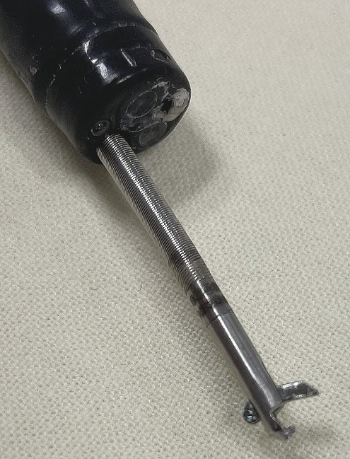
Figure 7. The SutuArt through-the-scope endoscopic needle holder. This figure is cited from [140].
EHS is capable of securing defect closure with high safety and strength. Therefore, it is a suitable candidate for large defects (measuring 20 mm to 30 mm or more) at various locations and depths. Consequently, it shows enormous potential in the field of advanced gastrointestinal endoscopic surgery [15, 74, 75]. However, the dissemination of this technique is limited by several practical problems, most prominently, the prolonged time required for the procedure and the technical complexity of EHS, which together necessitate a high degree of operator skill and a long period of training [74, 76, 77].
Recent advancements in endoscopic technology have given rise to new through-the-scope suturing devices. One of these innovations is the X-Tack (Apollo Endosurgery, Inc, Austin, Tex, USA), a through-the-scope continuous suturing device designed for efficient and secure closure of large or irregularly shaped GI defects (Figure 8) [78, 79]. The main parts of the X-Tack system comprise an applicator that converts the linear motion of the handle slider into rotational motion, four 5-mm-long surgical steel laser-cut helix tacks connected to the suture, and a cinch device used to tighten and secure the suture [80].

Figure 8. X-Tack suturing device. (A) X-Tack handle; (B) Pre-loaded helix tack. This figure is cited from [141].
During the procedure, the endoscopist uses the preloaded applicator to rotate and anchor the first helix tack into the tissue at the defect margin, thereby securing the initial end of the suture. The applicator catheter is then withdrawn and reloaded externally with the subsequent helical tacks, and the anchoring process is repeated at the defect edges. The subsequent helix tacks are preloaded on the suture and can slide along it. Therefore, a final cinch device is used to tighten the suture, thereby approximating the anchored helix tacks and achieving defect closure. After securing the other end of the suture with a T-tag deployed from the cinch device, the entire closure construct is stabilized, thus preventing re-opening and ensuring efficient and reliable closure [79].
Similar to EHS, X-Tack has been reported to have a high rate of clinical success [81]. However, as reported in a related study, several technical limitations may restrict its overall effectiveness [42]. For instance, difficulty in achieving full-thickness suturing results in a closure strength that is lower than that of TTSCs.
3.2 Over-the-scope suturing devices
As early as 1986, Swain et al. had already reported a chain-stitch suturing machine that was primarily made of rod-shaped transparent acrylic glass [82]. This suturing device is quite cumbersome and is fixed at the distal end of a double-channel endoscope. The tissue was drawn into the device’s chamber by the vacuum. Then a suture-loaded needle and a flexible control cable for the catch mechanism were carefully guided through the two instrument channels to perform the tissue approximation [82, 83].
In 1994, Swain et al. reported a tag-based suturing device used for fixing biosensors to the GI tract wall [84]. The two most significant features of this device were a specially designed tag and a hollow needle. In 1996, Kadirkamanathan et al. further developed a new endoscopic suturing device based on this principle for anti-reflux gastroplasty procedures [85]. This device connected the T-tag to a nylon thread which, after deployment from the hollow needle, was received by a dedicated reception chamber, thereby facilitating the continuation of the suturing process following external reloading [85]. Although the above devices were not developed with the direct purpose of suturing GI tract defects, the mechanical structures, suturing methods, and knot-tying techniques proposed during their development provided a reference for the research and development of future over-the-scope suturing devices.
Thereafter, the Apollo Group and Olympus Corporation designed prototype suturing devices that could be visualized endoscopically, the Eagle Claw series (Figure 9) [86]. Comprising a curved needle and opposable jaws, the devices were mounted on the exterior of the endoscopic distal end. The needle-tip of Eagle Claw II was non-detachable and had a hole for the thread to pass through. When the jaws closed, the tissue was punctured, and the thread passed through the tissue with the needle. A retrieval hook then retrieved one side of the suture through an extra channel provided by the device. After extracorporeal knotting, the knot was advanced back into the body using a second endoscope fitted with a knot-pushing cap [87]. The Eagle Claw V improved upon the previous generation by adding a detachable needle-tip with a thread and a catching cartridge attached to the other end of the suture, thereby avoiding extracorporeal knots [86]. When the detachable needle-tip, carrying the suture thread, punctured the tissue, it was captured by a catching cartridge [88]. Subsequent iterations of the device retained the basic mechanism and principle of the Eagle Claw V, featuring no significant alterations, and its feasibility and efficacy for hemostasis and gastrotomy closure were validated in a porcine gastric model [89, 90].

Figure 9. The eagle claw endoscopic suturing device. (A) Biological inspiration: curved talons; (B) Distal working end of the eagle claw device; (C) The complete system. This figure is cited from [141].
Building upon the Eagle Claw, the OverStitch (Apollo Endosurgery, Inc, Austin, Tex, USA) received Food and Drug Administration approval in 2008 after years of development [91]. The OverStitch is capable of safely and reliably achieving continuous suturing of the GI tract, enabling its use for endoscopic sleeve gastroplasty, full-thickness GI wall suturing, closure of post-ESD/EMR defects, management of fistulas or leaks, stent fixation, and various other indications [92-95]. It has become one of the most widely used over-the-scope suturing devices [96]. In contrast to its predecessors, the Eagle Claw series, OverStitch is a cap-based device, and is therefore more compact and maneuverable (Figure 10) [97]. This design eliminates the need for the large external catheter assembly attached to the outside of the endoscope body, thereby maximizing the inherent endoscopic flexibility [97]. However, its first and second iterations were only compatible with specific double-channel Olympus endoscopes, which consequently impeded its widespread clinical adoption [98].

Figure 10. Double-channel OverStitch suturing device with a tissue-capturing helix. (A) The needle driver handle mounted on the endoscope; (B) The Endcap mounted on the endoscopic distal end. This figure is cited from [141].
The continuous suturing mechanism of the OverStitch is engineered to emulate the operational mechanism of conventional manual suturing techniques for endoscopic application [91, 97]. The process begins as the unloaded needle driver closes and securely docks with the anchor exchange catheter within the instrument channel. The needle driver then reopens to retrieve the suture-attached anchor, thereby forming the functional operating unit. Following anchor retrieval, a helix device is deployed via the secondary instrument channel to capture and retract full-thickness tissue into the device’s jaw. Subsequently, closure of the needle driver advances the anchor to puncture and traverse the captured tissue. Upon full traversal, the deployed anchor is captured by the anchor exchange catheter, which simultaneously facilitates its separation from the needle driver, concluding the successful double full-thickness passage of the suture thread. Thereafter, the needle driver is reopened, and the suture thread, having passed through the tissue, is returned to the instrument channel, completing a single stitch. This entire sequence may then be repeated in vivo to establish a continuous running suture line. To secure the continuous suture line, the suture-attached anchor is released while the needle driver remains open, serving as the initial T-tag to fasten the distal end. In contrast, a dedicated cinch device is required proximally to tighten the suture thread and deploy a second T-tag for definitive fixation.
Compared to the OverStitch system, the OverStitch Sx (Apollo Endosurgery, Inc, Austin, Tex, USA) features two independent, external working channels that effectively substitute the two channels required by a double-channel endoscope (Figure 11) [99]. This innovative design significantly enhances device versatility by enabling compatibility with a wide range of standard single-channel flexible endoscopes, thereby promoting broader accessibility and application [99]. However, the addition of this external device may slightly restrict the overall flexibility and maneuverability of the endoscope [100].

Figure 11. Comparison of the OverStitch Sx and OverStitch endoscopic suturing systems. This figure is cited from [142].
The major drawback of regular TTSCs is their maximum opening width which is limited by the endoscopic working channel diameter. Whereas the clip span can be slightly increased by elongating the clip prongs, this physical limitation still considerably hampers their ability to close large mucosal or full-thickness defects. Compared with regular TTSCs, OTSCs provide a higher mechanical closure force, but they are not suitable for very large defects. Although endoscopic suturing devices can be used for larger defects, their higher economic cost and steep learning curve limit their widespread adoption in regular clinical practice.
As a result, over years of continuous clinical practice, endoscopists have come up with a multitude of advanced techniques to specifically surpass this limitation. These advanced clip-based closure techniques are systematically reviewed and presented in this paper. Depending on the main auxiliary instruments used for defect approximation, this review distinguishes clip-and-snare closure methods, loop-clip closure methods, clip-only closure methods, and clip-and-line closure methods.
4.1 Clip-and-snare closure methods
Clip-and-snare methods make use of regular clips in combination with detachable snares or similar auxiliary devices to achieve defect closure. In this approach, the method exerts continuous radial tension on the defect edges, which leads to the effective approximation of large defects.
The first use of a clip and snare for defect closure, in combination with a double-channel colonoscope, was reported by Matsuda et al. in 2004 [101]. An endoloop (Olympus, Tokyo, Japan) was placed around the defect, and regular clips were used to fix the two ends of the snare separately to the margins of the defect. Subsequent tightening of the endoloop provided the required radial tension for preliminary defect closure.
By inventing the endoscopic purse-string suture (EPSS) method in 2014, Zhang et al. significantly advanced the clip-and-snare closure method (Figure 12) [102]. The method involved placing multiple clips around the edges of the defect to fix the endoloop. The edges of the defect were drawn together by tightening the snare, and the method was confirmed effective for closing gastric wall defects less than 40 mm in diameter [102].

Figure 12. Schematic illustration of the EPSS procedure for closure of a gastrointestinal defect. (A, B) An initial clip is applied and an endoloop is positioned at the proximal margin of the defect; (C) Additional clips are deployed to secure the endoloop along the proximal edge; (D) The defect margins are approximated by tightening the endoloop; (E) Complete closure of the defect is achieved. This figure is cited from [102]. EPSS, endoscopic purse-string suture.
The initial dependence on a double-channel endoscope to allow simultaneous handling of both instruments limited the widespread adoption of the clip-and-snare method. However, Nomura et al. described an innovative single-channel endoscope method in 2018 [103]. Firstly, the operation begins with fastening the separated endoloop to one side of the clip using a surgical thread. The clip-connected endoloop is then withdrawn into the clip’s outer sheath, passed through the endoscope channel to the defect site, deployed from the sheath, and fixed to one side of the defect. The closure is completed using the standard purse-string steps.
After the EPSS had been established, endoscopists made various alterations to the technique. For example, placing the endoloop and the clips as a preventive measure before performing EFTR helps the subsequent closure of the created defect, thereby shortening the procedure time [104, 105]. Additionally, dental floss can be used for clip traction to prevent the clip from inverting during endoloop tightening. Alternatively, an additional endoloop can be placed over the EPSS for circumferential bundling and securing of all clips, thus effectively closing defects that EPSS cannot adequately address [106-108].
For more secure closure, Nishiyama et al. developed a novel technique named endoscopic ligation with O-ring closure (Figure 13) [109, 110]. In this method, the surgical nylon loop used for clip tightening replaces the detachable snare, and the endoscopic variceal ligation device is employed to gather the clips [109]. This process is then repeated to achieve total closure of large defects.

Figure 13. Schematic illustration of the endoscopic ligation with O-ring closure procedure. This figure is cited from [109].
4.2 Loop-clip closure methods
Loop-clip closure methods use clips that are either custom-made or commercially available and feature an integrated loop structure to achieve preliminary closure of large defects. Although terminology varies across the literature, this review refers to these devices collectively as “loop-clips”.
By way of an example in 2007, Fujii et al. utilized a regular clip to grasp a specially designed figure-of-8-shaped metallic loop, advancing it through the instrument channel to close a post-EMR defect (Figure 14) [111]. This method laid the foundation for subsequent loop-clip development. In 2008, Sakamoto et al. first described the loop-clip as a new closure device, which involved both a metallic and a nylon loop [112]. The operation of loop-clip methods is inherently simpler and more convenient than clip-and-snare methods. The process involves deploying the loop-clip at the defect margin, subsequently inserting a regular clip through the instrument channel to grasp the loop. The loop is then pulled to the opposing normal mucosa and the second clip is deployed, thus achieving preliminary closure of the large defect [113].

Figure 14. EMR and subsequent closure of the resulting mucosal defect using a figure-of-8-shaped metallic loop in combination with two resolution clips. This figure is cited from [111]. EMR, endoscopic mucosal resection.
The loop structure is highly versatile, with its material often consisting of metal, nylon, or rubber [111, 112, 114]. This variability accommodates diverse clinical scenarios, such as the closure method with an elastic ring reported by Kowazaki et al., which utilizes a clip with an attached elastic loop and also belongs to this category [114]. Regardless of the specific designs, the loop-clip essentially helps to turn a large defect into several smaller ones, which can then be closed individually with regular clips. In recent years, the loop-clip has been utilized to raise the mucosal layer and provide traction to the lesion, thus visually exposing more of the tissue that the endoscopist can dissect during ESD operations [115-117].
The most notable difference between loop-clip and clip-and-snare closure methods lies in how they apply force. Loop-clip closure methods use the loop as a medium. A second clip grips the loop of the first-deployed loop-clip and pulls it toward the opposing mucosa, thereby creating the traction. On the other hand, clip-and-snare closure methods utilize the loop structure itself, which is fastened to the defect rims by several clips. The radial contraction force resulting from loop tightening thus brings the defect rims toward the center.
4.3 Clip-only closure methods
Clip-only closure methods refer to techniques for closing large defects using only multiple clips, without auxiliary devices or tools. The double-layered closure method for closing large mucosal defects after ESD and EMR was introduced by Tanaka et al. in 2012 [118]. They deploy a set of clips along the long axis of the defect in the submucosal layer, thus reducing the defect area, and then, another set of clips is interwoven in the mucosal layer to achieve complete defect closure [118]. In 2017, Banno introduced the mucosa–submucosal clip closure method [119]. The principle involves orienting a regular clip parallel to the defect’s short axis, using its two prongs to hook the mucosa and submucosa separately, and then deploying it [119]. The process is carried out incrementally along the defect edge to bring the mucosal margins closer together, thus reducing the defect area that is finally closed with clips. Subsequent investigations, however, revealed that this method was limited to defects smaller than 50 mm and to those that were technically challenging [120, 121].
In 2018, the clip-on-clip closure method was proposed as an effective, straightforward, and safe technique (Figure 15) [122, 123]. First of all, a clip is deployed on the normal mucosa outside the defect circumference. Then, a second clip is placed on the first clip’s stem. To finish, one prong of the third clip is inserted into the space between the two prongs of the second clip, pulled to the opposite mucosa, and released. After confirming stability, additional clips are deployed to fully close the mucosal defect. In principle, the clip-on-clip closure method is functionally analogous to loop-clip methods, because the second clip is used to establish the traction bridge instead of a loop structure. Notably, to date, there have been no direct comparative ex vivo or clinical studies among different types of clip-only closure methods.

Figure 15. Schematic illustration of the CCCM. (A) A first clip is deployed on normal mucosa adjacent to the defect margin; (B) A second clip is attached to the stem of the first clip; (C, D) A third clip is advanced with one prong passing through the space between the jaws of the second clip to serve as a traction point; (E) The third clip is then secured to the contralateral normal mucosa to approximate the defect margins; (F) Complete closure is achieved with CCCM and additional clips. This figure is cited from [123]. CCCM, lip-on-clip closure method.
4.4 Clip-and-line closure methods
Clip-and-line closure methods achieve defect closure through the synergistic action of regular clips and a line, typically a suture or dental floss. This technique relies on linear tension exerted on the line, causing the clips anchored at the defect margin to converge, thereby achieving tissue approximation.
In 2016, Yamasaki et al. reported the line-assisted complete closure (LACC) method [124]. This method is effective for closing mucosal defects and perforations, significantly reducing the incidence of post-endoscopic submucosal dissection coagulation syndrome [125]. LACC is performed with only one line and regular clips. After binding the line to one clip prong, the clip is deployed on the side of the defect. A second clip is subsequently used to grasp the line and deploy it on the opposite side. If more clips are needed, a zigzag pattern is used along the margin [126]. Continuous tension is then applied to the line, approximating the defect for final closure with additional regular clips. Further research has found that LACC can be a viable option for large perforations and defects [127-129].
In 2021, Nomura et al. introduced the reopenable-clip over the line method (ROLM), which employs a line and reopenable clips to close defects without creating submucosal dead space [130, 131]. Compared to LACC, in ROLM the suture for the second and subsequent clips must be threaded extracorporeally through the tooth hole of the reopenable clip before insertion through the instrument channel [132]. This design ensures the line and the clip remain continuously connected, which eliminates the need for challenging intraprocedural line grasping and provides a more robust and stable connection for subsequent tension and defect approximation. It is important to note that both LACC and ROLM require the use of additional clips for complete closure of the defect after manual line tensioning, as the defect will reopen if tension is released. Furthermore, completion of the entire procedure necessitates the endoscopic transection of the line, which is typically performed using specialized accessories such as loop cutters or scissor forceps.
To overcome this limitation, Nomura et al. successfully combined ROLM with a modified locking-clip technique in 2022 [133]. The locking-clip technique is a technique that uses special TTSCs with specific mechanical structures [134]. The clip’s root is where the line is pre-threaded. When the clip is fully opened, the root of the clip and the line become firmly engaged. Therefore, endoscopists can first use ROLM with regular clips to gradually reduce defect area and approximate the tissue, and then they can apply modified locking-clip technique to tighten the line and fix it to the surrounding normal mucosa. After this important anchoring step, the line can be manually severed, thus allowing it to remain in vivo even under continuous high tension [133, 135].
The advancements in endoscopic closure devices, driven by clinician-engineer collaboration, have successfully transformed post-procedural GI defect management. Current innovation can be broadly categorized into two major directions: TTSCs and OTSCs and highly accurate instruments intended to imitate surgical suturing techniques. Within this landscape, the strategic use of TTSCs in combination with auxiliary devices effectively bypasses the mechanical constraints of single instruments, broadening their clinical applicability. Nonetheless, optimal clinical utilization demands a tailored approach, where endoscopists must integrate established procedural guidelines with defect complexity to judiciously select the most appropriate single device or combined closure strategy. Looking forward, although present closure devices, such as clips and emerging suturing systems, have shown functionality in various GI defects, the direction of future technological development should be that of enhanced reliability, operability, cost-effectiveness, and general ease of use of next-generation endoscopic closure devices.
Author contributions
Yuxiao Li contributed to the conceptualization and design of the review, performed the comprehensive literature search and data collection, prepared all tables, and was responsible for writing and revising the entire manuscript as the first author; Junjie Shen and Yuxuan Hou contributed to the literature review, data organization, and revision of sections of the manuscript; Shilong Li assisted with the literature screening, data verification, and proofreading of the manuscript for clarity and accuracy; Chengli Song provided critical feedback on the structure and content, and critically revised the manuscript before submission; Lin Mao, as the corresponding author, provided the overall project concept and methodology guidance, validated the findings, and provided final critical review and editing.
Funding
This research received no external funding.
Data availability
Not applicable.
Ethics approval and consent to participate
Not applicable.
Consent for publication
Not applicable.
Competing interests
The authors declare that they have no competing interests.
Acknowledgements
Not applicable.
[1] Ponsky JL, Strong AT. A history of flexible gastrointestinal endoscopy. Surg Clin North Am. 2020 Dec;100(6):971-992. https://doi.org/10.1016/j.suc.2020.08.013
[2] Rashid MU, Alomari M, Afraz S, Erim T. EMR and ESD: Indications, techniques and results. Surg Oncol. 2022 Aug;43:101742. https://doi.org/10.1016/j.suronc.2022.101742
[3] Pimentel-Nunes P, Libanio D, Bastiaansen BAJ, Bhandari P, Bisschops R, Bourke MJ, et al. Endoscopic submucosal dissection for superficial gastrointestinal lesions: European Society of Gastrointestinal Endoscopy (ESGE) Guideline - Update 2022. Endoscopy. 2022 Jun;54(6):591-622. https://doi.org/10.1055/a-1811-7025
[4] Inoue H, Maydeo A. Peroral endoscopic myotomy (POEM) opens the door of third-space endoscopy. Endoscopy. 2019 Nov;51(11):1010-1012. https://doi.org/10.1055/a-1019-1865
[5] Wang K, Gao P, Cai M, Song B, Zhou P. Endoscopic full-thickness resection, indication, methods and perspectives. Dig Endosc. 2023 Jan;35(2):195-205. https://doi.org/10.1111/den.14474
[6] Cecinato P, Sinagra E, Laterza L, Pianigiani F, Grande G, Sassatelli R, et al. Endoscopic removal of gastrointestinal lesions by using third space endoscopy techniques. Best Pract Res Cl Ga. 2024 Aug;71:101931. https://doi.org/10.1016/j.bpg.2024.101931
[7] Oka S, Tanaka S, Kanao H, Ishikawa H, Watanabe T, Igarashi M, et al. Current status in the occurrence of postoperative bleeding, perforation and residual/local recurrence during colonoscopic treatment in Japan. Dig Endosc. 2010 Oct;22(4):376-380. https://doi.org/10.1111/j.1443-1661.2010.01016.x
[8] Lee JH, Kedia P, Stavropoulos SN, Carr-Locke D. AGA clinical practice update on endoscopic management of perforations in gastrointestinal tract: Expert review. Clin Gastroenterol Hepatol. 2021 Nov;19(11):2252-2261.e2252. https://doi.org/10.1016/j.cgh.2021.06.045
[9] Dalal I, Andalib I. Advances in endoscopic resection: A review of endoscopic submucosal dissection (ESD), endoscopic full thickness resection (EFTR) and submucosal tunneling endoscopic resection (STER). Transl Gastroenterol Hepatol. 2022 Apr 25;7:19. https://doi.org/10.21037/tgh-2020-10
[10] Yoshida N, Wakabayashi N, Kanemasa K, Sumida Y, Hasegawa D, Inoue K, et al. Endoscopic submucosal dissection for colorectal tumors: Technical difficulties and rate of perforation. Endoscopy. 2009 Sep 10;41(9):758-761. https://doi.org/10.1055/s-0029-1215028
[11] Fujihara S, Mori H, Kobara H, Nishiyama N, Matsunaga T, Ayaki M, et al. Management of a large mucosal defect after duodenal endoscopic resection. World J Gastroenterol. 2016 Aug 7;22(29):6595-6609. https://doi.org/10.3748/wjg.v22.i29.6595
[12] Sun H, Cao T, Zhang F, Tao K, Xu H. Gastric defect closure after endoscopic full-thickness resection: The closing while dissecting technique. Surg Endosc. 2023 Jan;37(1):234-240. https://doi.org/10.1007/s00464-022-09457-7
[13] Paspatis GA, Arvanitakis M, Dumonceau JM, Barthet M, Saunders B, Turino SY, et al. Diagnosis and management of iatrogenic endoscopic perforations: European Society of Gastrointestinal Endoscopy (ESGE) Position Statement - Update 2020. Endoscopy. 2020 Sep;52(9):792-810. https://doi.org/10.1055/a-1222-3191
[14] Zhang Q, Han B, Xu J, Gao P, Shen Y. Clip closure of defect after endoscopic resection in patients with larger colorectal tumors decreased the adverse events. Gastrointest Endosc. 2015 Nov;82(5):904-909. https://doi.org/10.1016/j.gie.2015.04.005
[15] Spychalski M, Orzeszko Z, Kasprzyk P. Advancements in endoscopic closure: Embracing a new era of managing complications and postprocedural defects after endoscopic submucosal dissection. World J Gastrointest Endosc. 2025 Oct 16;17(10):107840. https://doi.org/10.4253/wjge.v17.i10.107840
[16] Granata A, Martino A, Ligresti D, Zito FP, Amata M, Lombardi G, et al. Closure techniques in exposed endoscopic full-thickness resection: Overview and future perspectives in the endoscopic suturing era. World J Gastrointest Surg. 2021 Jul 27;13(7):645-654. https://doi.org/10.4240/wjgs.v13.i7.645
[17] Kobara H, Tada N, Fujihara S, Nishiyama N, Masaki T. Clinical and technical outcomes of endoscopic closure of postendoscopic submucosal dissection defects: Literature review over one decade. Dig Endosc. 2023 Jan;35(2):216-231. https://doi.org/10.1111/den.14397
[18] Staudenmann D, Choi KKH, Kaffes AJ, Saxena P. Current endoscopic closure techniques for the management of gastrointestinal perforations. Ther Adv Gastrointest Endosc. 2022 Feb 27;15:26317745221076705. https://doi.org/10.1177/26317745221076705
[19] Medas R, Rodrigues-Pinto E. Technical review on endoscopic treatment devices for management of upper gastrointestinal postsurgical leaks. Gastroenterol Res Pract. 2023 Jun 12;2023(1):9712555. https://doi.org/10.1155/2023/9712555
[20] Papaefthymiou A, Norton B, Telese A, Murray C, Murino A, Johnson G, et al. Endoscopic suturing and clipping devices for defects in the GI tract. Best Pract Res Cl Ga. 2024 Jun;70:101915. https://doi.org/10.1016/j.bpg.2024.101915
[21] Hayashi T, Yonezawa M, Kuwabara T, kudoh I. The study on stanch clip for the treatment by endoscopy. Gastrointest Endosc. 1975 Feb 20;17(1):92-101. https://doi.org/10.11280/gee1973b.17.92
[22] Gong R, Wang S, Song J, He Z, Li P, Zhang S, et al. Closure methods for large defects after gastrointestinal endoscopic submucosal dissection. J Gastroenterol Hepatol. 2024 Dec;39(12):2511-2521. https://doi.org/10.1111/jgh.16722
[23] Conway JD, Adler DG, Diehl DL, Farraye FA, Kantsevoy SV, Kaul V, et al. Endoscopic hemostatic devices. Gastrointest Endosc. 2009 May;69(6):987-996. https://doi.org/10.1016/j.gie.2008.12.251
[24] McCarty TR, Jirapinyo P. Endoscopic closure: Tools and techniques. Gastrointest Endosc Clin N Am. 2023 Jan;33(1):169-182. https://doi.org/10.1016/j.giec.2022.08.003
[25] Wang TJ, Aihara H, Thompson AC, Schulman AR, Thompson CC, Ryou M. Choosing the right through-the-scope clip: A rigorous comparison of rotatability, whip, open/close precision, and closure strength (with videos). Gastrointest Endosc. 2019 Jan;89(1):77-86.e71. https://doi.org/10.1016/j.gie.2018.07.025
[26] Shin DY, Park S, Kim A, Kim ES, Jeon HH. Compatibility of endoclips in the gastrointestinal tract with magnetic resonance imaging. Sci Rep. 2020 Oct 6;10(1):16537. https://doi.org/10.1038/s41598-020-73726-5
[27] Hajiro K, Matsui H, Tsujimura D. Endoscopic hemostasis with hemoclips, local injection and other new techniques: The Japanese experience. Endoscopy. 1986;18:65-67. https://doi.org/10.1055/s-2007-1018433
[28] Hachisu T. Evaluation of endoscopic hemostasis usingan improved clipping apparatus. Surg Endosc. 1988 Mar;2(1):13-17. https://doi.org/10.1007/BF00591392
[29] Hachisu T, Miyazaki S, Hamaguchi K. Endoscopic clip-marking of lesions using the newly developed HX-3L clip. Surg Endosc. 1989 Sep;3(3):142-147. https://doi.org/10.1007/BF00591360
[30] Hachisu T, Yamada H, Satoh SI, Kouzu T. Endoscopic clipping with a new rotatable clip-device and a long clip. Dig Endosc. 1996 Apr;8(2):127-133. https://doi.org/10.1111/j.1443-1661.1996.tb00428.x
[31] Sonthalia N, Puri R, Sethi S. Role of clips, devices and sprays in endoscopic hemostasis: An update (with videos). Indian J Gastroenterol. 2025 Oct;44(5):595-604. https://doi.org/10.1007/s12664-025-01815-2
[32] Technology Assessment Committee, Chuttani R, Barkun A, Carpenter S, Chotiprasidhi P, Ginsberg GG, Hussain N, et al. Endoscopic clip application devices. Gastrointest Endosc. 2006 May;63(6):746-750. https://doi.org/10.1016/j.gie.2006.02.042
[33] Daram SR, Tang S, Wu R, To SD. Benchtop testing and comparisons among three types of through-the-scope endoscopic clipping devices. Surg Endosc. 2013 May;27(5):1521-1529. https://doi.org/10.1007/s00464-012-2679-4
[34] Romagnuolo J. Endoscopic clips: Past, present and future. Can J Gastroenterol. 2009 Mar;23(3):158-160. https://doi.org/10.1155/2009/515937
[35] Lin H. Can the triclip be successfully applied in clinical practice? Endoscopy. 2007 Apr;39(4):371. https://doi.org/10.1055/s-2007-966201
[36] Shinozaki S, Sunada K, Otake T, Lefor AT, Yamamoto H. Utility of a new reusable clip device for endoscopic treatment. Dig Endosc. 2015 Jan;27(1):106-112. https://doi.org/10.1111/den.12329
[37] Narihiro S, Yoshida M, Ohdaira H, Sato T, Suto D, Hoshimoto S, et al. A novel fluorescent marking clip for laparoscopic surgery of colorectal cancer: A case report. Int J Surg Case Rep. 2019;64:170-173. https://doi.org/10.1016/j.ijscr.2019.10.024
[38] Kato H, Kobayashi M, Yano M. Temporary sutures using reeponable clips and barbed threads. Dig Endosc. 2023 Jan;35(1):e18-19. https://doi.org/10.1111/den.14459
[39] Takeuchi Y, Shigita K, Asayama N, Aoyama T, Fukumoto A, Mukai S, et al. Clinical usefulness of the hold-and-drag closure using the SB clip for large mucosal defects after colorectal endoscopic submucosal dissection. Surg Endosc. 2023 Jul;37(7):5719-5725. https://doi.org/10.1007/s00464-023-10146-2
[40] Raju GS, Ahmed I, Xiao S, Brining D, Poussard A, Tarcin O, et al. Controlled trial of immediate endoluminal closure of colon perforations in a porcine model by use of a novel clip device (with videos). Gastrointest Endosc. 2006 Dec;64(6):989-997. https://doi.org/10.1016/j.gie.2006.06.058
[41] Verlaan T, Ponds FA, Bastiaansen BA, Bredenoord AJ, Fockens P. Single clips versus multi-firing clip device for closure of mucosal incisions after peroral endoscopic myotomy (POEM). Endosc Int Open. 2016 Oct;4(10):E1052-E1056. https://doi.org/10.1055/s-0042-113126
[42] Walradt T, Szvarca D, Zarrella S, Ryou M. New generation endoscopic closure devices for full-thickness defects: A comparative pre-clinical study (with videos). Surg Endosc. 2025 Jan;39(1):649-656. https://doi.org/10.1007/s00464-024-11434-1
[43] Raju GS. Endoscopic clip closure of gastrointestinal perforations, fistulae, and leaks. Dig Endosc. 2014 Jan;26(S1):95-104. https://doi.org/10.1111/den.12191
[44] Paspatis GA, Dumonceau JM, Barthet M, Meisner S, Repici A, Saunders BP, et al. Diagnosis and management of iatrogenic endoscopic perforations: European Society of Gastrointestinal Endoscopy (ESGE) Position Statement. Endoscopy. 2014 Aug;46(8):693-711. https://doi.org/10.1055/s-0034-1377531
[45] Rogalski P, Daniluk J, Baniukiewicz A, Wroblewski E, Dabrowski A. Endoscopic management of gastrointestinal perforations, leaks and fistulas. World J Gastroenterol. 2015 Oct 7;21(37):10542-10552. https://doi.org/10.3748/wjg.v21.i37.10542
[46] Zhang Q, Wang Z, Bai Y. A novel through-the-scope twin endoclip for a large mucosal closure in a live pig model. Endoscopy. 2019 Dec;51(12):E372-E373. https://doi.org/10.1055/a-0948-5252
[47] Zhang Q, Jin H, Shen Z, Ma X, Li C, Tang Z, et al. Novel through-the-scope twin clip for the closure of GI wounds: The first experimental survival study in pigs (with videos). Gastrointest Endosc. 2021 Oct;94(4):850-858.e2. https://doi.org/10.1016/j.gie.2021.04.027
[48] Shiomi D, Tanabe M, Uragami N, Toshimori A, Kawamata N, Muraoka M, et al. Clinical utility of a novel anchor pronged clip for mucosal defect closure after colorectal endoscopic submucosal dissection (with video). Endosc Int Open. 2024 Oct 10;12(10):E1127-E1133. https://doi.org/10.1055/a-2409-3622
[49] Inada T, Sumida Y, Matsumoto T, Fukuda SI, Maehara K, Akihiro H, et al. Muscle inversion closure technique with novel clips minimizes submucosal dead space after colorectal endoscopic submucosal dissection. Endoscopy. 2025 Dec;57(S 01):E717-E718. https://doi.org/10.1055/a-2612-3542
[50] Baron TH, Song LM, Ross A, Tokar JL, Irani S, Kozarek RA. Use of an over-the-scope clipping device: Multicenter retrospective results of the first U.S. experience (with videos). Gastrointest Endosc. 2012 Jul;76(1):202-208. https://doi.org/10.1016/j.gie.2012.03.250
[51] Kirschniak A, Kratt T, Stuker D, Braun A, Schurr MO, Konigsrainer A. A new endoscopic over-the-scope clip system for treatment of lesions and bleeding in the GI tract: First clinical experiences. Gastrointest Endosc. 2007 Jul;66(1):162-167. https://doi.org/10.1016/j.gie.2007.01.034
[52] Schurr MO, Arezzo A, Ho CN, Anhoeck G, Buess G, Di Lorenzo N. The OTSC clip for endoscopic organ closure in NOTES: Device and technique. Minim Invasiv Ther Allied Technol. 2008;17(4):262-266. https://doi.org/10.1080/13645700802275114
[53] Voermans RP, van Berge Henegouwen MI, Bemelman WA, Fockens P. Novel over-the-scope clip system for gastrotomy closure in natural orifice transluminal endoscopic surgery (NOTES): An ex vivo comparison study. Endoscopy. 2009 Dec;41(12):1052-1055. https://doi.org/10.1055/s-0029-1215231
[54] Renteln DV, Schmidt A, Vassiliou MC, Rudolph HU, Gieselmann M, Caca K. Endoscopic closure of large colonic perforations using an over-the-scope clip: A randomized controlled porcine study. Endoscopy. 2009 Jun;41(6):481-486. https://doi.org/10.1055/s-0029-1214759
[55] Armellini E, Crino SF, Orsello M, Ballare M, Tari R, Saettone S, et al. Novel endoscopic over-the-scope clip system. World J Gastroenterol. 2015 Dec 28;21(48):13587-13592. https://doi.org/10.3748/wjg.v21.i48.13587
[56] Monkemuller K, Toshniwal J, Zabielski M, Vornbrock K, Neumann H. Utility of the "bear claw", or over-the-scope clip (OTSC) system, to provide endoscopic hemostasis for bleeding posterior duodenal ulcers. Endoscopy. 2012;44(S 02):E412-E413. https://doi.org/10.1055/s-0032-1325737
[57] Weiland T, Fehlker M, Gottwald T, Schurr MO. Performance of the OTSC System in the endoscopic closure of iatrogenic gastrointestinal perforations: A systematic review. Surg Endosc. 2013 Jul;27(7):2258-2274. https://doi.org/10.1007/s00464-012-2754-x
[58] Bartell N, Bittner K, Kaul V, Kothari TH, Kothari S. Clinical efficacy of the over-the-scope clip device: A systematic review. World J Gastroenterol. 2020 Jun 28;26(24):3495-3516. https://doi.org/10.3748/wjg.v26.i24.3495
[59] Goenka MK, Rodge GA, Tiwary IK. Endoscopic management with a novel over-the-scope Padlock clip system. Clin Endosc. 2019 Nov;52(6):574-580. https://doi.org/10.5946/ce.2019.122
[60] Repici A, Arezzo A, De Caro G, Morino M, Pagano N, Rando G, et al. Clinical experience with a new endoscopic over-the-scope clip system for use in the GI tract. Dig Liver Dis. 2009 Jun;41(6):406-410. https://doi.org/10.1016/j.dld.2008.09.002
[61] Dinelli M, Omazzi B, Andreozzi P, Zucchini N, Redaelli A, Manes G. First clinical experiences with a novel endoscopic over-the-scope clip system. Endosc Int Open. 2017 Mar;5(3):E151-E156. https://doi.org/10.1055/s-0043-101692
[62] Kobara H, Mori H, Fujihara S, Nishiyama N, Chiyo T, Yamada T, et al. Outcomes of gastrointestinal defect closure with an over-the-scope clip system in a multicenter experience: An analysis of a successful suction method. World J Gastroenterol. 2017 Mar 7;23(9):1645-1656. https://doi.org/10.3748/wjg.v23.i9.1645
[63] Matthes K, Jung Y, Kato M, Gromski MA, Chuttani R. Efficacy of full-thickness GI perforation closure with a novel over-the-scope clip application device: An animal study. Gastrointest Endosc. 2011 Dec;74(6):1369-1375. https://doi.org/10.1016/j.gie.2011.07.057
[64] Singhal S, Changela K, Papafragkakis H, Anand S, Krishnaiah M, Duddempudi S. Over the scope clip: Technique and expanding clinical applications. J Clin Gastroenterol. 2013 Oct;47(9):749-756. https://doi.org/10.1097/MCG.0b013e318296ecb9
[65] von Renteln D, Schmidt A, Vassiliou MC, Gieselmann M, Caca K. Natural orifice transluminal endoscopic surgery gastrotomy closure with an over-the-endoscope clip: A randomized, controlled porcine study (with videos). Gastrointest Endosc. 2009 Oct;70(4):732-739. https://doi.org/10.1016/j.gie.2009.03.010
[66] Kobara H, Mori H, Nishiyama N, Fujihara S, Okano K, Suzuki Y, et al. Over-the-scope clip system: A review of 1517 cases over 9 years. J Gastroenterol Hepatol. 2019 Jan;34(1):22-30. https://doi.org/10.1111/jgh.14402
[67] Haito-Chavez Y, Law JK, Kratt T, Arezzo A, Verra M, Morino M, et al. International multicenter experience with an over-the-scope clipping device for endoscopic management of GI defects (with video). Gastrointest Endosc. 2014 Oct;80(4):610-622. https://doi.org/10.1016/j.gie.2014.03.049
[68] Schmidt A, Gölder S, Goetz M, Meining A, Lau J, von Delius S, et al. Over-the-scope clips are more effective than standard endoscopic therapy for patients with recurrent bleeding of peptic ulcers. Gastroenterology. 2018 Sep;155(3):674-686.e6. https://doi.org/10.1053/j.gastro.2018.05.037
[69] Ikeda K, Fritscher-Ravens A, Mosse CA, Mills T, Tajiri H, Swain CP. Endoscopic full-thickness resection with sutured closure in a porcine model. Gastrointest Endosc. 2005 Jul;62(1):122-129. https://doi.org/10.1016/S0016-5107(05)00517-1
[70] Sumiyama K, Gostout CJ, Rajan E, Bakken TA, Deters JL, Knipscheid MA. Endoscopic full-thickness closure of large gastric perforations by use of tissue anchors. Gastrointest Endosc. 2007 Jan;65(1):134-139. https://doi.org/10.1016/j.gie.2006.01.050
[71] Sporn E, Miedema BW, Bachman SL, Astudillo JA, Loy TS, Calaluce R, et al. Endoscopic colotomy closure after full thickness excision: Comparison of T fastener with multiclip applier. Endoscopy. 2008 Jul;40(7):589-594. https://doi.org/10.1055/s-2008-1077377
[72] Guarner-Argente C, Cordova H, Martinez-Palli G, Navarro-Ripoll R, Rodriguez-d'Jesús A, de Miguel CR, et al. Gastrotomy closure with a new tissue anchoring device: A porcine survival study. World J Gastroenterol. 2011 Apr 7;17(13):1732-1738. https://doi.org/10.3748/wjg.v17.i13.1732
[73] Goto O, Sasaki M, Ishii H, Horii J, Uraoka T, Takeuchi H, et al. A new endoscopic closure method for gastric mucosal defects: Feasibility of endoscopic hand suturing in an ex vivo porcine model (with video). Endosc Int Open. 2014 Jun 24;2(2):E111-E116. https://doi.org/10.1055/s-0034-1377180
[74] Niu C, Zhang J, Farooq U, Zahid S, Okolo PI. Endoscopic suturing for mucosal defect closure following endoscopic submucosal dissection: Systematic review and meta-analysis. Endosc Int Open. 2024 Oct 10;12(10):E1150-E1159. https://doi.org/10.1055/a-2411-8724
[75] Orzeszko Z, Kasprzyk P, Zawada U, Szura M, Spychalski M. Endoscopic hand suturing after advanced endoscopic procedures: Early outcomes of 31 cases in the upper and lower gastrointestinal tract (with video). Wideochir Inne Tech Maloinwazyjne. 2025 Feb 10;20(1):44-50. https://doi.org/10.20452/wiitm.2025.17928
[76] Scheppach MW, Nagl S, Muzalyova A, Classen J, Messmann H, Ebigbo A. Feasibility of a new endoscopic suturing device: A first Western experience (with video). Gastrointest Endosc. 2025 Jan;101(1):207-212. https://doi.org/10.1016/j.gie.2024.08.001
[77] Uozumi T, Abe S, Mizuguchi Y, Sekiguchi M, Toyoshima N, Takamaru H, et al. Endoscopic hand suturing using a modified through-the-scope needle holder for mucosal closure after colorectal endoscopic submucosal dissection: Prospective multicenter study (with video). Dig Endosc. 2024 Nov;36(11):1245-1252. https://doi.org/10.1111/den.14808
[78] Mahmoud T, Wong Kee Song LM, Stavropoulos SN, Alansari TH, Ramberan H, Fukami N, et al. Initial multicenter experience using a novel endoscopic tack and suture system for challenging GI defect closure and stent fixation (with video). Gastrointest Endosc. 2022 Feb;95(2):373-382. https://doi.org/10.1016/j.gie.2021.10.018
[79] Hernandez-Lara A, Garcia Garcia de Paredes A, Rajan E, Storm AC. Step-by-step instruction: Using an endoscopic tack and suture device for gastrointestinal defect closure. VideoGIE. 2021 Apr 5;6(6):243-245. https://doi.org/10.1016/j.vgie.2021.02.015
[80] Hernandez A, Marya NB, Sawas T, Rajan E, Gades NM, Wong Kee Song LM, et al. Gastrointestinal defect closure using a novel through-the-scope helix tack and suture device compared to endoscopic clips in a survival porcine model (with video). Endosc Int Open. 2021 Apr;9(4):E572-E577. https://doi.org/10.1055/a-1370-9256
[81] Suresh Kumar VC, Singh S, Loganathan P, Mohan BP, Aswath G, Akbar Khan HM, et al. Comparing the safety and efficacy of over-the-scope suturing, through-the-scope suturing, and endoscopic hand suturing for closure of GI defects after endoscopic resection: Systematic review and meta-analysis. Gastrointest Endosc. 2025 Sep;102(3):326-336.e325. https://doi.org/10.1016/j.gie.2025.03.002
[82] Swain CP, Mills TN. An endoscopic sewing machine. Gastrointest Endosc. 1986 Feb;32(1):36-38. https://doi.org/10.1016/s0016-5107(86)71727-6
[83] Swain CP. Endoscopic sewing and stapling machines. Endoscopy. 1997 Mar;29(3):205-210. https://doi.org/10.1055/s-2007-1004165
[84] Swain CP, Brown GJ, Gong F, Mills TN. An endoscopically deliverable tissue-transfixing device for securing biosensors in the gastrointestinal tract. Gastrointest Endosc. 1994 Nov-Dec;40(6):730-734. https://doi.org/10.1016/S0016-5107(94)70117-2
[85] Kadirkamanathan SS, Evans DF, Gong F, Yazaki E, Scott M, Swain CP. Antireflux operations at flexible endoscopy using endoluminal stitching techniques: An experimental study. Gastrointest Endosc. 1996 Aug;44(2):133-143. https://doi.org/10.1016/S0016-5107(96)70130-X
[86] Hu B, Chung SC, Sun LC, Lau JY, Kawashima K, Yamamoto T, et al. Developing an animal model of massive ulcer bleeding for assessing endoscopic hemostatic devices. Endoscopy. 2005 Sep;37(9):847-851. https://doi.org/10.1055/s-2005-870226
[87] Hu B, Chung SC, Sun LC, Kawashima K, Yamamoto T, Cotton PB, et al. Eagle Claw II: A novel endosuture device that uses a curved needle for major arterial bleeding: A bench study. Gastrointest Endosc. 2005 Aug;62(2):266-270. https://doi.org/10.1016/S0016-5107(05)00375-5
[88] Hu B, Chung SC, Sun LC, Lau JY, Kawashima K, Yamamoto T, et al. Endoscopic suturing without extracorporeal knots: A laboratory study. Gastrointest Endosc. 2005 Aug;62(2):230-233. https://doi.org/10.1016/S0016-5107(05)00331-7
[89] Chiu PW, Hu B, Lau JY, Sun LC, Sung JJ, Chung SS. Endoscopic plication of massively bleeding peptic ulcer by using the Eagle Claw VII device: A feasibility study in a porcine model. Gastrointest Endosc. 2006 Apr;63(4):681-685. https://doi.org/10.1016/j.gie.2005.10.030
[90] Chiu PW, Lau JY, Ng EK, Lam CC, Hui M, To KF, et al. Closure of a gastrotomy after transgastric tubal ligation by using the Eagle Claw VII: A survival experiment in a porcine model (with video). Gastrointest Endosc. 2008 Sep;68(3):554-559. https://doi.org/10.1016/j.gie.2008.03.1110
[91] ASGE Technology Committee, Banerjee S, Barth BA, Bhat YM, Desilets DJ, Gottlieb KT, et al. Endoscopic closure devices. Gastrointest Endosc. 2012 Aug;76(2):244-251. https://doi.org/10.1016/j.gie.2012.02.028
[92] Pauli EM, Delaney CP, Champagne B, Stein S, Marks JM. Safety and effectiveness of an endoscopic suturing device in a human colonic treat-and-resect model. Surg Innov. 2013 Dec;20(6):594-599. https://doi.org/10.1177/1553350613479204
[93] Stavropoulos S, Modayil R, Friedel D. Current applications of endoscopic suturing. World J Gastrointest Endosc. 2015 Jul 10;7(8):777-789. https://doi.org/10.4253/wjge.v7.i8.777
[94] Abu Dayyeh BK, Bazerbachi F, Vargas EJ, Sharaiha RZ, Thompson CC, Thaemert BC, et al. Endoscopic sleeve gastroplasty for treatment of class 1 and 2 obesity (MERIT): A prospective, multicentre, randomised trial. Lancet. 2022 Aug 6;400(10350):441-451. https://doi.org/10.1016/S0140-6736(22)01280-6
[95] Callahan ZM, Su B, Kuchta K, Conaty E, Novak S, Linn J, et al. Endoscopic suturing results in high technical and clinical success rates for a variety of gastrointestinal pathologies. J Gastrointest Surg. 2020 Feb;24(2):278-287. https://doi.org/10.1007/s11605-019-04485-6
[96] Maselli R, Palma R, Traina M, Granata A, Juzgado D, Bisello M, et al. Endoscopic suturing for GI applications: Initial results from a prospective multicenter European registry. Gastrointest Endosc. 2022 Nov;96(5):780-786. https://doi.org/10.1016/j.gie.2022.06.004
[97] Moran EA, Gostout CJ, Bingener J. Preliminary performance of a flexible cap and catheter-based endoscopic suturing system. Gastrointest Endosc. 2009 Jun;69(7):1375-1383. https://doi.org/10.1016/j.gie.2009.01.032
[98] Kantsevoy SV. The development of the overstitch system and its potentials. Gastrointest Endosc Clin N Am. 2020 Jan;30(1):107-114. https://doi.org/10.1016/j.giec.2019.08.004
[99] Keihanian T, Othman MO. OverStitch Sx Endoscopic suturing system in minimally invasive endoscopic procedures: Overview of its safety and efficacy and comparison to overstitch™. Expert Rev Med Devices. 2022 Jan;19(1):11-23. https://doi.org/10.1080/17434440.2022.2019579
[100] Chou CK, Chen SS, Lee CY, Toh DE, Chuah YY, Tsai KF, et al. Comprehensive defect closure with overStitch Sx after endoscopic resection of gastric and duodenal subepithelial lesions. J Formos Med Assoc. 2025 Oct 28;S0929-6646(25)00470-X. https://doi.org/10.1016/j.jfma.2025.09.005
[101] Matsuda T, Fujii T, Emura F, Kozu T, Saito Y, Ikematsu H, et al. Complete closure of a large defect after EMR of a lateral spreading colorectal tumor when using a two-channel colonoscope. Gastrointest Endosc. 2004 Nov;60(5):836-838. https://doi.org/10.1016/S0016-5107(04)02033-4
[102] Zhang Y, Wang X, Xiong G, Qian Y, Wang H, Liu L, et al. Complete defect closure of gastric submucosal tumors with purse-string sutures. Surg Endosc. 2014 Jun;28(6):1844-1851. https://doi.org/10.1007/s00464-013-3404-7
[103] Nomura T, Kobayashi M, Morikawa T, Horiki N. Clip-fixed endoloop: An efficacious new method for mucosal defect closure. Endoscopy. 2018 Jun;50(6):E126-E127. https://doi.org/10.1055/s-0044-101025
[104] Wu N, Liu S, Chen M, Zeng X, Wang F, Zhang J, et al. The preprune-string suture technique for gastric defect after endoscopic full-thickness resection (with video). Medicine (Baltimore). 2018 Sep;97(36):e12118. https://doi.org/10.1097/MD.0000000000012118
[105] Li M, Zhang Y, Sun F, Huai M, Zhang F, Qu C, et al. Feasibility and efficacy of endoscopic purse-string suture-assisted closure for mucosal defects induced by endoscopic manipulations. World J Gastroenterol. 2023 Jan 28;29(4):731-743. https://doi.org/10.3748/wjg.v29.i4.731
[106] Li B, Huang S, Liao S, He M, Xiao Y, Zhong E, et al. Modified endoscopic purse-string suture with dental floss traction in the management of a duodenal defect. Endoscopy. 2025 Dec;57(S 01):E491-E492. https://doi.org/10.1055/a-2590-8450
[107] Wang S, Ji M, Huang X, Li Y, Yu S, Zhao Y, et al. Double-nylon purse-string suture in closing postoperative wounds following endoscopic resection of large (≥ 3 cm) gastric submucosal tumors. World J Gastroenterol. 2024 Mar 7;30(9):1143-1153. https://doi.org/10.3748/wjg.v30.i9.1143
[108] Walia A, Trasolini RP, Shahidi N. Double-nylon purse-string suture technique: Another addition to the endoscopist's toolbox for full-thickness defect closure. World J Gastroenterol. 2024 Jul 7;30(25):3152-3154. https://doi.org/10.3748/wjg.v30.i25.3152
[109] Nishiyama N, Kobara H, Kobayashi N, Chiyo T, Fujihara S, Yachida T, et al. Novel endoscopic ligation with O-ring closure involving muscle layer of a gastric artificial defect. Endoscopy. 2020 Nov;52(11):E413-E414. https://doi.org/10.1055/a-1149-8597
[110] Nishiyama N, Kobara H, Kobayashi N, Chiyo T, Tada N, Kozuka K, et al. Efficacy of endoscopic ligation with O-ring closure for prevention of bleeding after gastric endoscopic submucosal dissection under antithrombotic therapy: A prospective observational study. Endoscopy. 2022 Nov;54(11):1078-1084. https://doi.org/10.1055/a-1782-3448
[111] Fujii T, Ono A, Fu KI. A novel endoscopic suturing technique using a specially designed so-called "8-ring" in combination with resolution clips (with videos). Gastrointest Endosc. 2007 Dec;66(6):1215-1220. https://doi.org/10.1016/j.gie.2007.05.054
[112] Sakamoto N, Beppu K, Matsumoto K, Shibuya T, Osada T, Mori H, et al. "Loop Clip", a new closure device for large mucosal defects after EMR and ESD. Endoscopy. 2008 Sep;40:E97-E98. https://doi.org/10.1055/s-2007-995604
[113] Mori H, Sakamoto N, Osada T, Ritsuno H, Hidaka Y, Nakae K, et al. The 'Loop Clip' is useful for closing large mucosal defects after colorectal endoscopic submucosal dissection: A preliminary clinical study. Dig Endosc. 2011 Oct;23(4):330-331. https://doi.org/10.1111/j.1443-1661.2010.01099.x
[114] Kowazaki Y, Fukuda H, Saito I, Sawano T, Kurokawa T, Kanzaki N, et al. A novel method for endoscopic closure of endoscopic submucosal dissection-induced defects in the colorectum: The closure method with an elastic-rubber ring (CMER). Endoscopy. 2021 Aug;54(08):E407-E408. https://doi.org/10.1055/a-1550-2246
[115] Ge PS, Aihara H. A novel clip-band traction device to facilitate colorectal endoscopic submucosal dissection and defect closure. VideoGIE. 2020 May 31;5(5):180-186. https://doi.org/10.1016/j.vgie.2020.01.012
[116] Jinushi R, Tashima T, Terada R, Miyaguchi K, Katsuda H, Ogawa T, et al. Effectiveness of a multi-loop traction device for colorectal endoscopic submucosal dissection performed by trainees: A pilot study. Scientific Reports. 2022 Jun 17;12(1):10197. https://doi.org/10.1038/s41598-022-14407-3
[117] Muramatsu T, Tashima T, Ishikawa T, Terada R, Kawasaki T, Itoi T, et al. A novel triangle traction method using a clip-band traction device for endoscopic submucosal dissection of early gastric cancer involving the pyloric ring. Endoscopy. 2022 Dec;55(S 01):E207-E208. https://doi.org/10.1055/a-1956-1349
[118] Tanaka S, Toyonaga T, Obata D, Ishida T, Morita Y, Azuma T. Endoscopic double-layered suturing: A novel technique for closure of large mucosal defects after endoscopic mucosal resection (EMR) or endoscopic submucosal dissection (ESD). Endoscopy. 2012;44(S 02):E153-E154. https://doi.org/10.1055/s-0031-1291673
[119] Banno S, Nishizawa T, Kinoshita S, Mori H, Uraoka T. Endoscopic mucosa-submucosa clip closure method. Endoscopy. 2017 Dec;49(12):E307-E308. https://doi.org/10.1055/s-0043-119349
[120] Kinoshita S, Nishizawa T, Fujimoto A, Mori H, Nakazato Y, Kikuchi M, et al. Efficacy of mucosa-submucosa clip closure method after gastric endoscopic submucosal dissection. World J Gastrointest Endosc. 2020 Jan 16;12(1):17-22. https://doi.org/10.4253/wjge.v12.i1.17
[121] Nishizawa T, Banno S, Kinoshita S, Mori H, Nakazato Y, Hirai Y, et al. Feasibility of endoscopic mucosa-submucosa clip closure method (with video). Endosc Int Open. 2018 Aug;6(8):E1070-E1074. https://doi.org/10.1055/a-0630-0566
[122] Nomura T, Kamei A, Sugimoto S, Oyamada J. New closure method for a mucosal defect after endoscopic submucosal dissection: The clip-on-clip closure method. Endoscopy. 2018 May;50(5):547-548. https://doi.org/10.1055/s-0044-100486
[123] Nomura T, Matsuzaki I, Sugimoto S, Oyamada J, Kamei A, Kobayashi M. Clip-on-clip closure method for a mucosal defect after colorectal endoscopic submucosal dissection: A prospective feasibility study. Surg Endosc. 2020 Mar;34(3):1412-1416. https://doi.org/10.1007/s00464-019-07195-x
[124] Yamasaki Y, Takeuchi Y, Kato M, Uedo N, Ishihara R. Line-assisted complete closure of large gastric mucosal defects by use of multiple clip-and-line technique. VideoGIE. 2016 Sep 3;1(3):49-50. https://doi.org/10.1016/j.vgie.2016.08.008
[125] Yamasaki Y, Takeuchi Y, Iwatsubo T, Kato M, Hamada K, Tonai Y, et al. Line-assisted complete closure for a large mucosal defect after colorectal endoscopic submucosal dissection decreased post-electrocoagulation syndrome. Dig Endosc. 2018 Sep;30(5):633-641. https://doi.org/10.1111/den.13052
[126] Ominami M, Nagami Y, Tanaka C, Sakai T, Fukunaga S, Watanabe T, et al. Endoscopic technique for closure of a large gastric tube perforation by using endoclips with line-assisted complete closure. Endoscopy. 2019 Mar;51(3):E49-E50. https://doi.org/10.1055/a-0800-8502
[127] Nakahira H, Takeuchi Y, Garcia JS, Morita Y, Uedo N, Ishihara R, et al. Line-assisted endoscopic complete closure of a large perforation during colonic endoscopic submucosal dissection. Endoscopy. 2018 Jan;50(1):E32-E33. https://doi.org/10.1055/s-0043-121137
[128] Yamasaki Y, Takeuchi Y, Uedo N, Hanaoka N, Higashino K, Ishihara R, et al. Line-assisted complete closure of duodenal mucosal defects after underwater endoscopic mucosal resection. Endoscopy. 2017 Feb;49(S 01):E37-E38. https://doi.org/10.1055/s-0042-120707
[129] Nakahira H, Takeuchi Y, Kanesaka T, Uedo N, Ishihara R. Wide-field underwater EMR followed by line-assisted complete closure for a large duodenal adenoma. VideoGIE. 2019 Oct;4(10):469-471. https://doi.org/10.1016/j.vgie.2019.07.004
[130] Nomura T, Sugimoto S, Tsuda N, Matsushima R, Oyamada J, Kamei A. Mucosal defect closure after duodenal endoscopic submucosal dissection using the reopenable-clip over the line method. JGH Open. 2021 Jul;5(7):831-833. https://doi.org/10.1002/jgh3.12577
[131] Nomura T, Sugimoto S, Temma T, Oyamada J, Ito K, Kamei A. Reopenable clip-over-the-line method for closing large mucosal defects following gastric endoscopic submucosal dissection: Prospective feasibility study. Dig Endosc. 2023 May;35(4):505-511. https://doi.org/10.1111/den.14466
[132] Nomura T, Sugimoto S, Temma T, Oyamada J, Ito K, Kamei A, et al. Gastric antral defect closure after endoscopic submucosal dissection with the reopenable clip-over-the-line method using a multibending scope. Endoscopy. 2023 Dec;55(S 01):E866-E867. https://doi.org/10.1055/a-2107-2474
[133] Nomura T, Sugimoto S, Kawabata M, Oyamada J, Kamei A. Large colorectal mucosal defect closure post-endoscopic submucosal dissection using the reopenable clip over line method and modified locking-clip technique. Endoscopy. 2022 Feb;54(2):E63-E64. https://doi.org/10.1055/a-1381-6435
[134] Nomura T, Kamei A, Sugimoto S, Harada T, Oyamada J. Thread-assisted mucosal defect closure using the locking-clip technique after gastric endoscopic submucosal dissection. Endoscopy. 2018 Dec;50(12):E342-E343. https://doi.org/10.1055/a-0725-7754
[135] Nomura T, Kamei A, Sugimoto S. Clip line traction method using locking clip technique for colorectal endoscopic submucosal dissection. Dig Endosc. 2019 May;31(3):e72-e73. https://doi.org/10.1111/den.13364
[136] Nabi Z, Reddy DN. Endoscopic closure of gastrointestinal defects: Devices and techniques. Expert Rev Gastroenterol Hepatol. 2025 Sep;19(9):973-983. https://doi.org/10.1080/17474124.2025.2539793
[137] Al-Bawardy B, Rajan E, Wong Kee Song LM. Over-the-scope clip-assisted endoscopic full-thickness resection of epithelial and subepithelial GI lesions. Gastrointest Endosc. 2017 May;85(5):1087-1092. https://doi.org/10.1016/j.gie.2016.08.019
[138] Wei MT, Ahn JY, Friedland S. Over-the-scope clip in the treatment of gastrointestinal leaks and perforations. Clin Endosc. 2021 Nov;54(6):798-804. https://doi.org/10.5946/ce.2021.250
[139] Guarner-Argente C, Córdova H, Martínez-Pallí G, Navarro R, Cuatrecasas M, Rodríguez de Miguel C, et al. Yes, we can: Reliable colonic closure with the Padlock-G clip in a survival porcine study (with video). Gastrointest Endosc. 2010 Oct;72(4):841-844. https://doi.org/10.1016/j.gie.2010.06.054
[140] Troya J, Fuchs KH, Hann A, Meining A. Technical aspects of endoscopic internal drainage procedure, secured by endoscopic suture fixation: Experimental study. Endosc Int Open. 2026 Jan 14;14:a27779441. https://doi.org/10.1055/a-2777-9441
[141] McKhann C, Lee LS. Saving patients one stitch at a time? iGIE. 2023 Jun;2(2):202-208. https://doi.org/10.1016/j.igie.2023.04.002
[142] Keihanian T, Zabad N, Khalaf M, Abdel-Jaber W, Kim YI, Jawaid SA, et al. Safety and efficacy of a novel suturing device for closure of large defects after endoscopic submucosal dissection (with video). Gastrointest Endosc. 2023 Sep;98(3):381-391. https://doi.org/10.1016/j.gie.2023.04.006
ISSN: 2957-5478
Volume 4, Issue 2
June 2026
Pages: 77-177


 Log In

 On This Page
 CITE
CITE