 Log In
Search




 Open Access
 csong@usst.edu.cn
A closed-loop regulation system utilizing peripheral nerves was developed to enhance motor control.
The COMSOL Multiphysics electric field model achieved millimeter-level spatial resolution and effective deep tissue penetration.
Experimental results confirmed the stability and reproducibility of the generated interference stimulation signals.
Deep modulation through peripheral pathways may reduce surgical risks and support future closed-loop Parkinson’s disease therapies.
Open Accesscsong@usst.edu.cnA closed-loop regulation system utilizing peripheral nerves was developed to enhance motor control.
The COMSOL Multiphysics electric field model achieved millimeter-level spatial resolution and effective deep tissue penetration.
Experimental results confirmed the stability and reproducibility of the generated interference stimulation signals.
Deep modulation through peripheral pathways may reduce surgical risks and support future closed-loop Parkinson’s disease therapies.
Keywords: Parkinson’s disease, Resting tremor, Temporal interference, Peripheral nerve, Electrical stimulation
Parkinson’s disease (PD) ranks just behind Alzheimer’s disease as the most common neurodegenerative disorder affecting humans. It is estimated that the number of PD patients in China reaches nearly 3 million, accounting for about 23% of the global PD patient population, with new cases increasing annually by approximately 100,000 [1]. The disease manifests through four primary motor dysfunctions, with tremors being one of the most prominent symptoms; 75% of patients experience resting tremors. The pathological focus of PD lies in the loss of dopaminergic neurons in the substantia nigra, leading to abnormal functioning of the ventral intermediate nucleus of thalamus [2]. The resting tremors, characterized by rhythmic movements of limbs at a frequency of 4–6 Hz, significantly disrupt patients’ daily lives and social interactions. Current therapies face several limitations. Pharmacological treatments often lead to drug resistance and side effects. Deep Brain Stimulation can restore functional activity in the basal ganglia by delivering electrical pulses but requires invasive electrode implantation, which carries surgical risks and substantial costs—averaging between $20,000 and $50,000 per patient. In contrast, Transcutaneous Electrical Nerve Stimulation is a non-invasive option typically costing around $50 to $200 for devices, but it requires higher electrical current intensities, which can induce muscle fatigue and discomfort, limiting its long-term effectiveness [3]. In 2017, Temporal Interference Stimulation (TIS) emerged as a promising non-invasive technique that utilizes the superposition of two high-frequency electric fields, producing an envelope-modulated low-frequency stimulation in deep brain areas. Unlike Deep Brain Stimulation, which primarily targets specific regions through invasive methods and is limited in adaptability once implanted, TIS can achieve millimeter-level precision while targeting multiple anatomical structures in a non-invasive manner, significantly reducing operational costs by avoiding surgical expenses [4-6]. The dual-directional coupling mechanism between the central and peripheral nervous systems is noteworthy: abnormal oscillatory signals in the basal ganglia-thalamic area can transmit through peripheral nerves to induce tremors, whereas peripheral electrical stimulation can help normalize basal ganglia activity via the ventral intermediate nucleus of thalamus [7, 8]. A closed-loop peripheral nerve stimulation scheme based on high-frequency carrier wave temporal interference was developed for peripheral nerve modulation via a closed-loop control system. COMSOL Multiphysics was used to simulate the bioelectric conduction model to verify these hypotheses. Different anatomical structures, such as the median nerve and radial nerve, can thus be precisely targeted with millimeter accuracy to generate biologically relevant electric field modulation with low-frequency periodic interference components using this method. This intervention methodology breaks through the spatial constraints of external stimulation from traditional electrical stimulation that limit the effectiveness of the stimulation and brings TIS theory into the field of peripheral nerve regulation for non-invasive and precise tremor intervention [9]. It is an indirect approach for controlling tremor symptoms through non-invasive external stimulation technology that is inexpensive and convenient for patients with Parkinsonian tremors [10-12].
2.1 Theoretical basis
According to Maxwell’s equations, for non-invasive electrical stimulation, the corresponding mathematical model satisfies:

In the above equation, σ represents the conductivity distribution in Ω, which is the domain of the stimulated electric field [13]. J is the external electrical stimulation applied through the electrodes, and u is the resultant potential distribution. After obtaining the potential distribution u by solving the aforementioned equations, the electric field E and current density J can be calculated based on the following definitions:

2.2 Principles of peripheral nerve electrical stimulation
Peripheral nerve electrical stimulation is based on the principle of neural plasticity, which activates neuronal activity by stimulating peripheral nerves in the human body, thereby influencing the conduction and regulation of neural signals [14]. This technique makes full use of the sensitivity of neurons to external stimuli, adjusting the transmission and response of neural signals to achieve therapeutic effects [14, 15].
Peripheral nerve electrical stimulation generates electrical pulses that act on the target nerves, causing the cell membrane to transition from a resting potential to an action potential. This results in changes in neuronal excitability, subsequently modulating neural networks activity and signal transmission. In patients with resting tremors, despite the integrity of the neural command conduction system, peripheral nerve electrical stimulation primarily targets the median and radial nerves with low-frequency pulse stimulation. This regulates the activity of the thalamic ventral intermediate nucleus, generating nerve impulses and correcting abnormal discharges, thereby restoring normal bodily function [2]. Furthermore, peripheral nerve electrical stimulation promotes nerve regeneration and functional recovery, improving patients’ muscle control and quality of life. The principle of peripheral nerve electrical stimulation is illustrated in Figure 1.


2.3 Principles of TIS
TIS applies multiple high-frequency (kHz range) sinusoidal alternating currents to the body surface, utilizing the frequency difference between different electrode pairs (e.g., Δf=10 Hz) to generate interference effects in deep tissues [3]. This mechanism is based on the principle of physical superposition. As high-frequency currents penetrate surface tissues, the frequency difference creates a low-frequency envelope-modulated electric field (with a frequency of Δf). Due to the higher response threshold of neurons to high-frequency electric fields, only the low-frequency envelope electric field produced by the deep tissue interference results in a depolarization response. By dynamically adjusting the electrode configuration, frequency difference, and current ratio, it is possible to precisely control the spatial location and depth of the stimulation target [4]. This three-phase mechanism of “high-frequency conduction - deep interference - low-frequency activation” overcomes the penetration depth limitations of traditional non-invasive neural modulation techniques, providing a non-invasive and highly targeted precision modulation solution for PD. The mechanism of TIS is shown in Figure 2.

2.4 Experimental materials and models
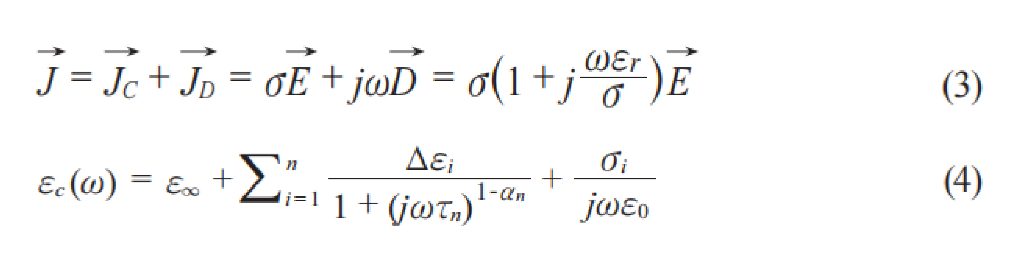
When traditional low-frequency and direct current stimulation satisfies the condition of Equation (3) 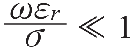 , the current density is solely dependent on the conductivity of human tissues, allowing for the neglect of displacement current [21]. However, under the influence of mid-frequency currents in the kHz range, the contribution of displacement current becomes significant due to the increased frequency and the high relative permittivity characteristics of human tissues. This results in a notable reduction in the total impedance of the tissues, facilitating an increase in overall current density and making it possible to stimulate nerves located deep within the body [22].
The dispersive characteristics of human tissues lead to frequency-dependent dielectric parameters (εr,σ), resulting in nonlinear variations in penetration depth for different frequency currents. To quantitatively assess the differences in penetration, a three-layer tissue model established in COMSOL (Figure 3) based on frequency-varying electrical characteristic parameters (Table 1) was employed, utilizing finite element methods to solve for the current density distribution within the body at different frequencies. Using the interpolation function module of COMSOL Multiphysics, the multidimensional electrical characteristic parameters from Table 1 were imported into the biological tissue material model, and a parametric scan was conducted to study the penetration of sinusoidal waves at different frequencies in human tissues.


Equation (3) represents Maxwell’s current conduction equation, and Equation (4) denotes the tissue dispersive equation.
Temporal interference employs two typical electrode configurations, as shown in Figure 4: a symmetric and a crossed configurations. In the simulation, to more accurately reflect real-world conditions, a dual-channel stimulation system with reverse current-driven isolation was used to effectively avoid ground loops and noise interference. The circuit model is illustrated in Figure 5.


3.1 Validation of neuronal low-pass characteristics
To verify the low-frequency nature of neurons, the researchers used the traditional Hodgkin-Huxley model and carried out simulations using NEURON 8.0 software.
During the simulation, pulse waves of different frequencies were applied to evaluate the response of the neuron membrane potential to different stimulation frequencies. The aim of this study is to explore the neurons’ discharge patterns and adaptability through the analysis of different stimulation frequencies.
The simulation outcomes, depicted in Figure 6, confirmed that neurons possess low-pass characteristics, which therefore serve as a theoretical basis for further research.

Figure 6 shows the results from electrophysiological simulation tests that rely on the Hodgkin-Huxley neuronal model. The results show that the firing pattern of neuronal action potentials is highly dependent on the frequency of the stimulating current. Stimulation based on the Hodgkin-Huxley model implemented in the NEURON simulation platform showed that when a positive pulse current exceeded the threshold, sodium channels were activated, resulting in a fast depolarization process. A negative current, on the other hand, leads to a hyperpolarization which is mainly caused by potassium ion efflux, after which the membrane potential is slowly reset to its original state by means of leakage currents.
The frequency-response relationship was very clear in the simulation data: within the 200 Hz stimulation limit, at least, the action potential firing frequency was linearly dependent on input current frequency. That is, the higher the stimulation frequency, the greater the firing rate. Nevertheless, when the stimulation frequency is raised to 1 kHz, neurons display remarkable low-pass filtering features, and thus the membrane potential stays at the resting level. The absence of neuronal responses to high-frequency stimulation provides a plausible explanation for the therapeutic application of TIS in peripheral nerves.
3.2 KHz current penetration simulation model
To quantitatively analyze the experimental results, a study was conducted on the current density distribution within the subcutaneous depth range of 0–20 mm along the vertical direction of the midplane of the three-layer simplified model (Figure 7). The results are shown in Figure 3.

3.3 Simulation of temporal interference on peripheral nerves
The optimal stimulation frequency range was narrowed down to 2–5 kHz, after a detailed factor analysis of previous studies on low-frequency characteristics and current penetration, as well as the work of Ward et al. [23]. This discovery gives vital parameter support to simulation research and hardware design of peripheral nerve electrical stimulation.
In the COMSOL simulation, the circuit model of Figure 5 was implemented, where two sets of electrodes were placed at arc positions around a circle with a radius of 4 cm—specifically at the 1/8, 3/8, 5/8, and 7/8 positions. Here, V1=V2=30 V, f1=2.0 kHz, and f2=2.04 kHz. The results (Figure 8) state that the symmetric electrode has a different distribution of envelope modulation amplitude across different directions. Along the y-axis, the amplitude distribution is less dispersed toward the deeper entities, and the modulation region assumes an elliptical shape. In contrast, the crossed electrode shows a more concentrated amplitude distribution in both the x and y directions, making it suitable for wide-area stimulation.

The Ey component of the point at the center of Figure 4 is plotted within 50 ms, as shown in Figure 9. As an example, points A (13.03, 3.74) and B (38.03, 3.74) were taken; the period is 0.04 s, and the frequency is 40 Hz, which corresponds to the envelope frequency of |f1-f2|. Moreover, the envelope impact range under six different electrode configurations was analyzed, and the results are given in Figure 10.


As shown in Figure 10, among the six representative electrode configurations, the symmetric configuration stands out as having superior positional accuracy with respect to the crossed configuration. Detailed comparison studies of the six-electrode configurations (Figure 10) revealed key performance differences: the symmetric configuration significantly exceeds the crossed configuration in positional accuracy, while the crossed configuration holds advantages in stimulation coverage and field uniformity. The symmetric electrode can provide targeted positioning with a spatial resolution of the order of millimeters by simply changing the electrode crossing angle and inter-electrode distance, which can be leveraged for more precise and controllable peripheral nerve stimulation based on neural modulation requirements.
After finishing the above simulations, a portable closed-loop dual-channel high-frequency Parkinson’s electrical stimulator was developed to bring precise relief to Parkinson’s patients through targeted electrical stimulation of their nervous system. However, the device requires further experimental validation to ascertain that it is indeed safe and effective and has good potential for clinical use. Figure 11 depicts the layout of the electrical stimulation system, while Figure 12 shows the power supply module printed circuit board layout.


This research represents the final stage of our work on a closed-loop regulation system based on TIS theory and utilizing peripheral nerve modulation effect. COMSOL Multiphysics was used to create a bioelectric model that accurately describes the low-frequency interference electric field resulting from two high-frequency carrier waves. The predicted spatial resolution of the projection field shows that it can map a very small target region, down to a millimeter scale. The electric field generated by the interference has the potential to penetrate both skin and soft tissues, resulting in stable and reliable interference signals.
Compared to existing technologies, this represents a significant advancement, as our method enables non-invasive deep modulation via peripheral nerve pathways. By avoiding the inherent risks associated with surgical implants, this technology offers a promising alternative for the treatment of tremors related to PD. Furthermore, the findings underscore the potential of the “external carrier - internal interference” mechanism to overcome the spatial limitations of traditional electrical stimulation methods. By finely tuning parameters such as phase, frequency, and amplitude, precise control over the electric field strength and distribution can be achieved, laying a critical foundation for the advancement of TIS technology toward wearable devices and intelligent closed-loop systems.
Based on its millimeter-level resolution, non-invasive deep modulation, and highly adjustable parameters, this technology may be miniaturized into portable or wearable devices. By integrating real-time physiological signal monitoring modules, a smart closed-loop system could be established. This system would enable real-time detection of the physiological state of tremors and dynamically adjust TIS stimulation parameters for adaptive, on-demand intervention. This not only provides a promising new solution for personalized and long-term management of tremors in patients with PD but also highlights the significant potential of the “external carrier - internal interference” mechanism in next-generation intelligent neural modulation devices.
Despite these positive outcomes, further in-depth analysis of the limitations is necessary. The simulation model incorporates several simplifying assumptions, such as tissue uniformity and isotropy, which may lead to deviations in the results. Specifically, the responses of human biological systems are highly complex, and inter-individual physiological differences have not been fully considered in the model. Variations in physiological conditions, skin thickness, tissue types, and pathological changes among different patients can all influence the propagation of the interference electric field and the efficacy of neural stimulation. It is important to recognize that further validation in clinical settings is required to evaluate the practical application value and biological safety of this innovative method. Future research should focus on conducting clinical trials to assess the effectiveness and safety of the closed-loop control system in real-world scenarios. Overall, this study lays the groundwork for the development of advanced non-invasive treatment devices aimed at PD patients and opens new pathways to improve clinical outcomes in tremor management.
Author contributions
Chengli Song contributed to the conception of the study; Chengli Song, Zhiyuan Zhao contributed significantly to analysis and manuscript preparation; Zhiyuan Zhao performed the data analyses and wrote the manuscript; Zhiyuan Zhao, Wenjie Yu helped perform the analysis with constructive discussions.
Funding
This research received no external funding.
Data availability
All data generated or analyzed during this study are included in this published article.
Ethics approval and consent to participate
Not applicable.
Consent for publication
All authors consent to the publication of this study.
Competing interests
The authors declare that they have no competing interests.
Acknowledgements
During the course of this research, we did not receive any form of financial support. All required funding was sourced personally by the researchers or their affiliated institutions. The design, implementation, data analysis, and writing of the conclusions of this study were not influenced by any funding sponsors.
[1] Qi S, Yin P, Wang L, Qu M, Kan G, Zhang H, et al. Prevalence of Parkinson’s disease: A community‐based study in China. Mov Disord. 2021 Dec;36(12):2940-2944. https://doi.org/10.1002/mds.28762
[2] Bloem BR, Okun MS, Klein C. Parkinson’s disease. Lancet. 2021 Jun 12;397(10291):2284-2303. https://doi.org/10.1016/S0140-6736(21)00218-X
[3] Pascual-Valdunciel A, Hoo GW, Avrillon S, Barroso FO, Goldman JG, Hernandez-Pavon JC, et al. Peripheral electrical stimulation to reduce pathological tremor: A review. J Neuroeng Rehabil. 2021 Feb 15;18(1):33. https://doi.org/10.1186/s12984-021-00811-9
[4] Grossman N, Bono D, Dedic N, Kodandaramaiah SB, Rudenko A, Suk HJ, et al. Noninvasive deep brain stimulation via temporally interfering electric fields. Cell. 2017 Jun 1;169(6):1029-1041.e16. https://doi.org/10.1016/j.cell.2017.05.024
[5] Mirzakhalili E, Barra B, Capogrosso M, Lempka SF. Biophysics of temporal interference stimulation. Cell Syst. 2020 Dec 16;11(6):557-572.e5. https://doi.org/10.1016/j.cels.2020.10.004
[6] Stoupis D, Samaras T. Non-invasive stimulation with temporal interference: optimization of the electric field deep in the brain with the use of a genetic algorithm. J Neural Eng. 2022 Sep 22;19(5):056018. https://doi.org/10.1088/1741-2552/ac89b3
[7] Dideriksen JL, Laine CM, Dosen S, Muceli S, Rocon E, Pons JL, et al. Electrical stimulation of afferent pathways for the suppression of pathological tremor. Front Neurosci. 2017 Apr 4;11:178. https://doi.org/10.3389/fnins.2017.00178
[8] Bisio A, Avanzino L, Gueugneau N, Pozzo T, Ruggeri P, Bove M. Observing and perceiving: a combined approach to induce plasticity in human motor cortex. Clin Neurophysiol. 2015 Jun;126(6):1212-1220. https://doi.org/10.1016/j.clinph.2014.08.024
[9] Liu R, Zhu G, Wu Z, Gan Y, Zhang J, Liu J, et al. Temporal interference stimulation targets deep primate brain. Neuroimage. 2024 May 1;291:120581. https://doi.org/10.1016/j.neuroimage.2024.120581
[10] Xiao Q, Zhong Z, Lai X, Qin H. A multiple modulation synthesis method with high spatial resolution for noninvasive neurostimulation. PLoS One. 2019 Jun 20;14(6):e0218293. https://doi.org/10.1371/journal.pone.0218293
[11] Qi S, Liu X, Yu J, Liang Z, Liu Y, Wang X. Temporally interfering electric fields brain stimulation in primary motor cortex of mice promotes motor skill through enhancing neuroplasticity. Brain Stimul. 2024 Mar-Apr;17(2):245-257. https://doi.org/10.1016/j.brs.2024.02.014
[12] Zhu Z, Yin L. A mini-review: Recent advancements in temporal interference stimulation in modulating brain function and behavior. Front Hum Neurosci. 2023 Sep 14;17:1266753. https://doi.org/10.3389/fnhum.2023.1266753
[13] Kim J, Wichmann T, Inan OT, Deweerth SP. A wearable system for attenuating essential tremor based on peripheral nerve stimulation. IEEE J Transl Eng Health Med. 2020 Apr 6;8:1-11. https://doi.org/10.1109/JTEHM.2020.2985058
[14] Pascual-Valdunciel A, Rajagopal A, Pons JL, Delp S. Non-invasive electrical stimulation of peripheral nerves for the management of tremor. J Neurol Sci. 2022 Apr 15;435:120195. https://doi.org/10.1016/j.jns.2022.120195
[15] Zhong Q, Zhu F. Trends in prevalence cases and disability-adjusted life-years of Parkinson’s disease: Findings from the global burden of disease study 2019. Neuroepidemiology. 2022;56(4):261-270. https://doi.org/10.1159/000524208
[16] Ghobadi-Azbari P, Jamil A, Yavari F, Esmaeilpour Z, Malmir N, Mahdavifar-Khayati R, et al. fMRI and transcranial electrical stimulation (tES): A systematic review of parameter space and outcomes. Prog Neuropsychopharmacol Biol Psychiatry. 2021 Apr 20;107:110149. https://doi.org/10.1016/j.pnpbp.2020.110149
[17] Evers J, Sridhar K, Liegey J, Brady J, Jahns H, Lowery M. Stimulation-induced changes at the electrode–tissue interface and their influence on deep brain stimulation. J Neural Eng. 2022 Jul 4;19(4):046004. https://doi.org/10.1088/1741-2552/ac7ad6
[18] Beliaeva V, Savvateev I, Zerbi V, Polania R. Toward integrative approaches to study the causal role of neural oscillations via transcranial electrical stimulation. Nat Commun. 2021 Apr 14;12(1):2243. https://doi.org/10.1038/s41467-021-22468-7
[19] Albouy P, Weiss A, Baillet S, Zatorre RJ. Selective entrainment of theta oscillations in the dorsal stream causally enhances auditory working memory performance. Neuron. 2017 Apr 5;94(1):193-206.e5. https://doi.org/10.1016/j.neuron.2017.03.015
[20] Albouy P, Baillet S, Zatorre RJ. Driving working memory with frequency‐tuned noninvasive brain stimulation. Ann N Y Acad Sci. 2018;1423(1):126-137. https://doi.org/10.1111/nyas.13664
[21] Yang C, Xu Y, Feng X, Wang B, Du Y, Wang K, et al. Transcranial temporal interference stimulation of the right globus pallidus in Parkinson’s disease. Mov Disord. 2025 Jun;40(6):1061-1069. https://doi.org/10.1002/mds.29967
[22] Vassiliadis P, Beanato E, Popa T, Windel F, Morishita T, Neufeld E, et al. Non-invasive stimulation of the human striatum disrupts reinforcement learning of motor skills. Nat Hum Behav. 2024 Aug;8(8):1581-1598. https://doi.org/10.1038/s41562-024-01901-z
[23] Ward AR, Robertson VJ. Variation in motor threshold with frequency using kHz frequency alternating current. Muscle Nerve. 2001 Oct;24(10):1303-1311. https://doi.org/10.1002/mus.1148
ISSN: 2957-5478
Volume 4, Issue 3
September 2026
Pages: 178-283


 Log In

 On This Page
 CITE
CITE