 Log In
Search




 Open Access
 whta01956@btch.edu.cnOpen Accesswhta01956@btch.edu.cn
Ischemia–reperfusion (IR) injury reflects secondary pathological processes arising after reperfusion of hypoperfused organs. The central paradox is that reperfusion itself drives much of the tissue damage. In the lungs, reperfusion-induced damage is associated with structural and functional impairment. This acute phase is characterized by excessive release of reactive oxygen species (ROS) and inflammatory mediators, with consequent loss of membrane integrity and dysregulation of immune responses, which can result in microvascular hyperpermeability (pulmonary edema) and acute lung injury. Mechanistically, the cathepsin C axis in alveolar macrophages is an important modulator of this process; cathepsin C has been shown to regulate p38 mitogen-activated protein kinase-mediated p47 phosphorylation for nicotinamide adenine dinucleotide phosphate oxidase activation and is associated with increased pro-inflammatory cytokines [1].
Beyond the lungs, the heart exhibits a similar paradoxical response. This cascade, driven by ROS-mediated oxidative stress, advances toward proteomic modifications which compromise membrane integrity and provoke programmed cell death (apoptosis) and contribute to myocyte loss. Importantly, IR is characterized by the selective release of lysine crotonylation-modified proteins—predominantly from the contractile apparatus—with kinetic profiles that correlate with injury severity and suggest autonomous cross-reactivity of post-translational modifications. Despite this, there remains a significant gap in our understanding of the mechanisms at play and the exact molecular signatures that can be exploited for precision intervention.
Underlying these organ-specific manifestations is a common molecular hub: iron dysregulation. ROS generation is the initiator, with the buildup of unstable Fe(II) as a central molecular hub. The overwhelming intracellular Fe(II), in addition to directly oxidizing membrane structures, lipids, and nuclear DNA, also elicits a systemic inflammatory cascade. Interestingly, iron overload inhibits [4Fe-4S] cluster biogenesis, leading to the destabilization of lipoic acid synthase. As a result, the depletion of [4Fe-4S] inhibits protein lipoylation—a metabolic contributor to IR injury in the kidney [2]. Thus, exploring how Fe(II) interacts with the mitochondrial cofactors provides a theoretical basis to identify specific drugs against reperfusion injury in clinical practice.
2.1 The interplay of ferroptosis, apoptosis, and necrosis
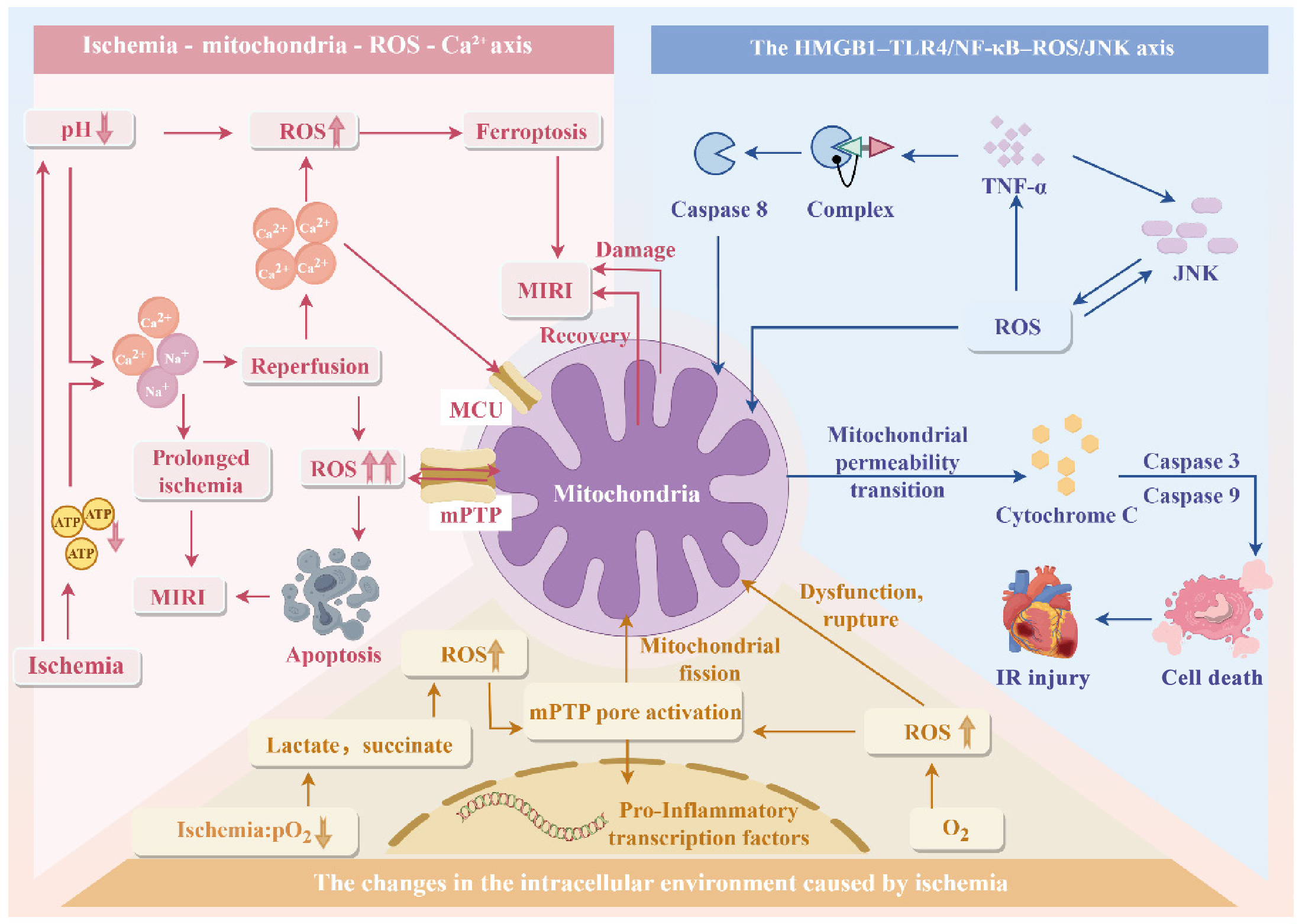
Iron-overload-activated ferroptosis is an important contributor to cell death in IR injury. Ischemia produces an energetic catastrophe, while reperfusion triggers a synergistic cascade—manifesting as bursts of ROS generation and inflammatory signaling, along with Ca2+ dysregulation—that acutely augments cellular demise. The ALOX15–15-HpETE axis, involving arachidonate 15-lipoxygenase and its metabolite 15-hydroperoxyeicosatetraenoic acid, is specifically involved in cardiomyocyte pathology. The pathological outcome is determined by a balance between regulated apoptosis and uncontrolled necrosis. Apoptosis is immunologically “silent”, whereas gross tissue injury tends to yield necrosis. This transition liberates damage-associated molecular patterns, generating a positive feedback loop of tertiary inflammation and tissue destruction.
2.2 Initiation and persistence of the inflammatory cascade
Overwhelming inflammation is the major pathogenetic mechanism in IR. The major immunological stimuli that recruit immune effectors to the affected organ are fluctuations in oxygen levels and metabolic substrates after reperfusion, which trigger the release of damage-associated molecular patterns and cytokines. These leukocytes release ROS and proteases, which induce a self-perpetuating inflammatory loop and promote lesion expansion. Li et al. found that NOD-like receptor family pyrin domain containing 3 inflammasome activation and astrocyte pyroptosis after cerebral IR are dependent on the lipocalin 2–24p3 receptor axis [3]. This neurodegeneration-promoting autocrine and paracrine feedback is accelerated in the final stages of programmed cell death, when functional recovery becomes unattainable.
2.3 The mitochondrial–redox nexus: From bioenergetic failure to cell death

3.1 Myocardial IR injury: Clinical insights and therapeutic innovations
3.2 Neurological sequelae of reperfusion injury
The neurological sequelae of reperfusion injury appear as a secondary event after restoration of blood flow to the brain. This process is characterized by the rapid disruption of cellular homeostasis; although flow is restored, neurons often irreversibly transition into a state of metabolic and structural dysfunction mediated by an intense inflammatory response. One important contributor to this neuroinflammatory cascade is the HDAC3–p65–cGAS–STING signaling pathway, which can potentiate innate immunity and contribute to focal neurological deficits [5]. In addition, disrupted neurotransmitter release in the damaged areas prevents synaptic stability, resulting in a loss of function. Finally, cerebral reperfusion injury not only amplifies the primary ischemic insult but also triggers sustained—and in many cases permanent—neurological dysfunction, which constitutes a critical barrier to clinical recovery.
3.3 Challenges of reperfusion injury in organ transplantation
4.1 Preconditioning: Priming the endogenous defense
4.2 Pharmacological modulation of reperfusion injury
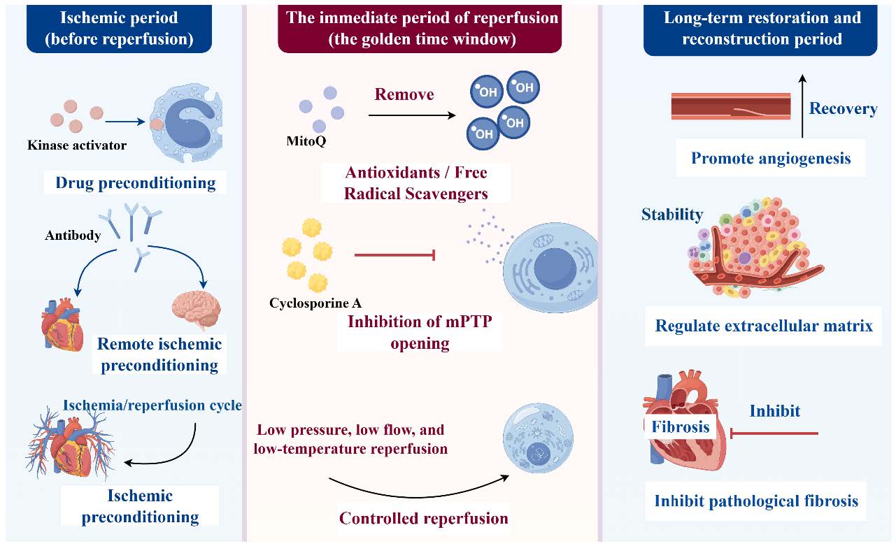
Pharmacological approaches to reperfusion injury focus mainly on oxidative stress, inflammatory pathways, and programmed cell death. Ligustilide (LIG) is an emerging candidate with potent therapeutic efficacy by virtue of its dual antioxidant and anti-inflammatory properties. LIG exerts cytoprotective effects by inhibiting ROS bursts, limiting inflammatory responses, and preventing microvascular perfusion loss. In cerebral IR models, LIG protects mitochondrial function and protects against both apoptosis and necrosis. Mechanistically, these effects are mediated by crosstalk with the PTEN-induced kinase 1 axis, which both prevents functional deterioration and promotes recovery. As such, a stratified phase-based intervention paradigm is proposed, targeting tissue resilience during ischemia, ROS scavenging and mitochondrial stabilization during reperfusion, and structural remodeling modulation during prolonged recovery (Figure 2).
4.3 Frontiers in targeted precision therapy
Targeted interventions have also emerged as a focus of IR research. One such agent is complement receptor 2–complement receptor 1-related protein y (CR2–Crry), a molecular species well-positioned for targeted intervention. CR2–Crry is a high-affinity, domain-specific inhibitor of the complement cascade that constrains pathogenic immune activation and thereby prevents tissue injury. CR2–Crry provides strong local protection with limited systemic coverage when administered prophylactically, a beneficial feature in the context of brain death-induced IR injury potentiation [8]. Indeed, the significant free radical scavenging activity, Hedgehog signaling inhibition, and anti-inflammatory properties of this compound suggest broad applicability across a spectrum of CV and cerebrovascular disorders. In fact, CR2–Crry is a prototype of the vectorial specificity needed to modulate reperfusion injuries that can best be applied using a site-directed mechanism of action and where expectations for a high-efficacy, low-toxicity paradigm can be realized.
Current biomarkers for IR injury have notable limitations, prompting the search for more sensitive and early indicators. The most widely used circulating biomarkers include lactate dehydrogenase and creatine kinase-MB, which are commonly applied to support the clinical assessment of IR injury. In this context, experimental modeling—including studies of epigenetic and metabolic remodeling—demonstrates systemic alterations in the levels of these enzymes, reflecting membrane disruption and leakage of cytosolic proteins, thereby contributing to their diagnostic relevance in cardiac injury assessment [9]. There is a shift away from traditional diagnostic markers toward mechanism-based biomarkers and next-generation multi-parameter panels. Therefore, future studies should explore ultra-early injury biomarkers with high sensitivity, such as microRNAs, selective post-translational modifications, or changes in metabolic intermediates that allow quantification of therapeutic effects.
Another promising direction is research on solute carrier family 26 member 6 (Slc26a6), which highlights its importance in modulating the IR environment. This mechanism has two components: modulation of acid-base balance in the myocardium via the Cl-/HCO3- exchanger, and modulation of ischemia-induced acidosis. It has been demonstrated that ablation of Slc26a6 protects against ischemic metabolic acidosis through elevation of myocardial pH. Importantly, a key role of Slc26a6 has been described in the regulation of transmembrane ionic balance, which is a major determinant of myocardial susceptibility to IR injury [10]. Elucidation of these complex Slc26a6 functions should augment our understanding of cardiovascular pathophysiology with potential relevance to novel therapeutic frontiers and interdisciplinary science.
Finally, inadequate blinding in trial designs represents a significant barrier to the translation of IR-based interventions. Trials lacking double-blinding—where either the experimenter or the subjects remain unblinded—are susceptible to experimenter bias and subject expectancy effects. These confounders act to cloud therapeutic assessment and mask the actual effect of therapies directed toward reperfusion. Thus, future studies should be double-blind randomized controlled trials with rigorous methodological quality and greater clinical relevance. Here, we emphasize the importance of a strict blinding process to ensure objectivity and accurate assessment of treatment effects, thereby facilitating the bench-to-bedside translation of IR therapies.
Author contributions
Haotian Wu contributed to manuscript writing and figure preparation; Huan Zhang designed the work; Siyi Yan supervised the work. All authors have read and approved the final manuscript.
Funding
This research received no external funding.
Data availability
Data sharing not applicable to this article as no datasets were generated or analyzed during the current study. All information is derived from publicly available articles and datasets.
Ethics approval and consent to participate
Not applicable. This manuscript does not contain any studies with human participants or animals performed by any of the authors.
Consent for publication
Not applicable. This manuscript does not include details, images, or videos relating to an individual person.
Competing interests
The authors declare that they have no competing interests.
Acknowledgements
Not applicable.
ISSN: 2957-5443
Volume 4, Issue 3
September 2026
Pages: 278-374


 Log In

 On This Page
 CITE
CITE