 Log In
Search




 Open Access
 yyh.8@163.com
Yaohua Yu, Department of Anesthesiology, The First Hospital of Putian, No. 449, Nanmen West Road, Chengxiang District, Putian 351100, Fujian, China. E-mail: yyh.8@163.com.
Received May 11, 2026; Accepted September 4, 2026; Published September 30, 2026
Open Accessyyh.8@163.comYaohua Yu, Department of Anesthesiology, The First Hospital of Putian, No. 449, Nanmen West Road, Chengxiang District, Putian 351100, Fujian, China. E-mail: yyh.8@163.com.
Received May 11, 2026; Accepted September 4, 2026; Published September 30, 2026
Keywords: Hip fracture, Severe cardiac disease, Ultrasound-guided anesthesia, Transthoracic echocardiography, Hemodynamic management
Elderly patients with hip fractures are frequently complicated by severe cardiac disease, resulting in a markedly increased risk of perioperative cardiac mortality and complications [1-3]. Anesthetic management requires a delicate balance between adequate analgesia and the maintenance of fragile hemodynamic stability [4,5]. With the widespread adoption of perioperative ultrasound, anesthesia practice is evolving from experience-based interventions toward precision-guided management [6,7]. This report presents four representative cases to illustrate how ultrasound techniques can be integrated into anesthesia strategies tailored to specific cardiac pathophysiology.
2.1 Case 1: Severe mitral stenosis—open reduction and internal fixation of left femoral fracture
A 66-year-old woman with a left intertrochanteric femoral fracture and a history of cerebral infarction was admitted. Echocardiography revealed severe mitral stenosis (valve area 0.77 cm2), pulmonary hypertension (85 mmHg, World Health Organization Group 2, Grade III), atrial fibrillation, and enlargement of the left atrium (69 mm) and right atrium (45 mm). N‑terminal pro‑brain natriuretic peptide (NT-proBNP) was 11,979 pg/mL. The relevant images are shown in Figure 1.

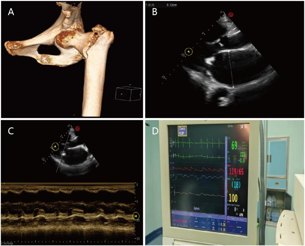
(B) Intraoperative TTE, parasternal longaxis view, showing left atrial enlargement (61 mm); (C) M-mode echocardiography of the mitral valve showing a ‘doming’ (hockey-stick–like) appearance; (D) Intraoperative monitoring revealing atrial fibrillation, with IBP of 119/65 mmHg and CVP of 18 mmHg. CT, computed tomography; TTE, transthoracic echocardiography; IBP, invasive arterial blood pressure; CVP, central venous pressure.
Anesthetic challenges: Maintaining adequate preload, reducing pulmonary artery pressure, ensuring right ventricular forward flow, controlling ventricular rate in atrial fibrillation, and preserving sufficient diastolic filling time.
Management: Ultrasound-guided low-dose combined spinal–epidural anesthesia (CSEA) was used, employing a long-axis out-of-plane approach in the right lateral position, with sensory block level controlled at T10. Invasive arterial blood pressure (IBP) and central venous pressure (CVP) were monitored. Ventricular rate was controlled preoperatively. A 300 mL crystalloid preload was administered before neuraxial anesthesia, and intraoperative fluid therapy followed a restrictive strategy, maintaining CVP at the upper normal limit. Sedation was achieved with dexmedetomidine (20 μg/h), and norepinephrine (0.05–0.1 μg/kg/min) was used to maintain blood pressure while avoiding tachycardia and hypotension.
Ultrasound application: Preoperative ultrasound-guided fascia iliaca compartment block (FICB) for positioning and postoperative analgesia; ultrasound-guided internal jugular vein and radial artery cannulation; ultrasound-guided CSEA using a long-axis out-of-plane approach; preoperative ultrasound assessment of cardiac chamber size, systolic function, and valvular status; intermittent transthoracic echocardiography (TTE) evaluation of right ventricular function, mitral valve motion, and fluid responsiveness.
Outcome: Surgery was completed successfully with stable hemodynamics and good postoperative recovery.
2.2 Case 2: Post-myocardial infarction status—posterior approach hemiarthroplasty
A 67-year-old man with a right femoral neck fracture, hypertension, and cardiorenal insufficiency presented with troponin I of 9.470 μg/L and NT-proBNP of 5,100 pg/mL. Electrocardiogram showed complete right bundle branch block and T-wave abnormalities suggestive of inferior ischemia. Echocardiography demonstrated interventricular septal thickening (15 mm) and left ventricular wall thickening (14 mm), left ventricular ejection fraction 60%, impaired diastolic function, and arrhythmia. Coronary computed tomography angiography and angiography revealed severe multivessel stenosis. The relevant images are shown in Figure 2.
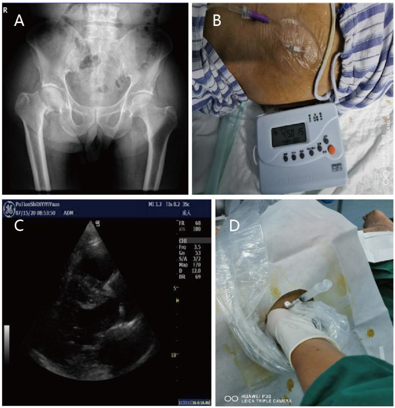
Anesthetic challenges: Maintaining hemodynamic stability, preventing perioperative myocardial ischemia, and managing antiplatelet therapy.
Management: After multidisciplinary consultation and medical optimization, troponin I decreased to 0.300 μg/L. Ultrasound-guided FICB combined with low-dose CSEA (T10 level) was performed. IBP and CVP were monitored. Heart rate and blood pressure were strictly maintained within ±20% of baseline to preserve myocardial oxygen supply–demand balance. Severe hypovolemia was avoided, and inotropes were used cautiously. Sedation with dexmedetomidine (16 μg/h) and norepinephrine infusion (0.05–0.1 μg/kg/min) maintained stable hemodynamics.
Ultrasound application: Continuous FICB for 7 days; ultrasound-guided vascular access and neuraxial anesthesia; preoperative assessment of ventricular wall thickness, chamber size, and systolic function; intraoperative TTE monitoring of left ventricular function and fluid responsiveness.
Outcome: Hemodynamics remained stable without ischemic events. Dual antiplatelet therapy was resumed postoperatively, and coronary stenting was performed six months later.
2.3 Case 3: Severe pulmonary hypertension—open reduction and internal fixation of right femoral fracture
An 82-year-old man with a right intertrochanteric fracture, chronic obstructive pulmonary disease, and cor pulmonale was evaluated. Echocardiography showed severe tricuspid regurgitation, pulmonary hypertension (94 mmHg), and impaired left ventricular diastolic function. The relevant images are shown in Figure 3.
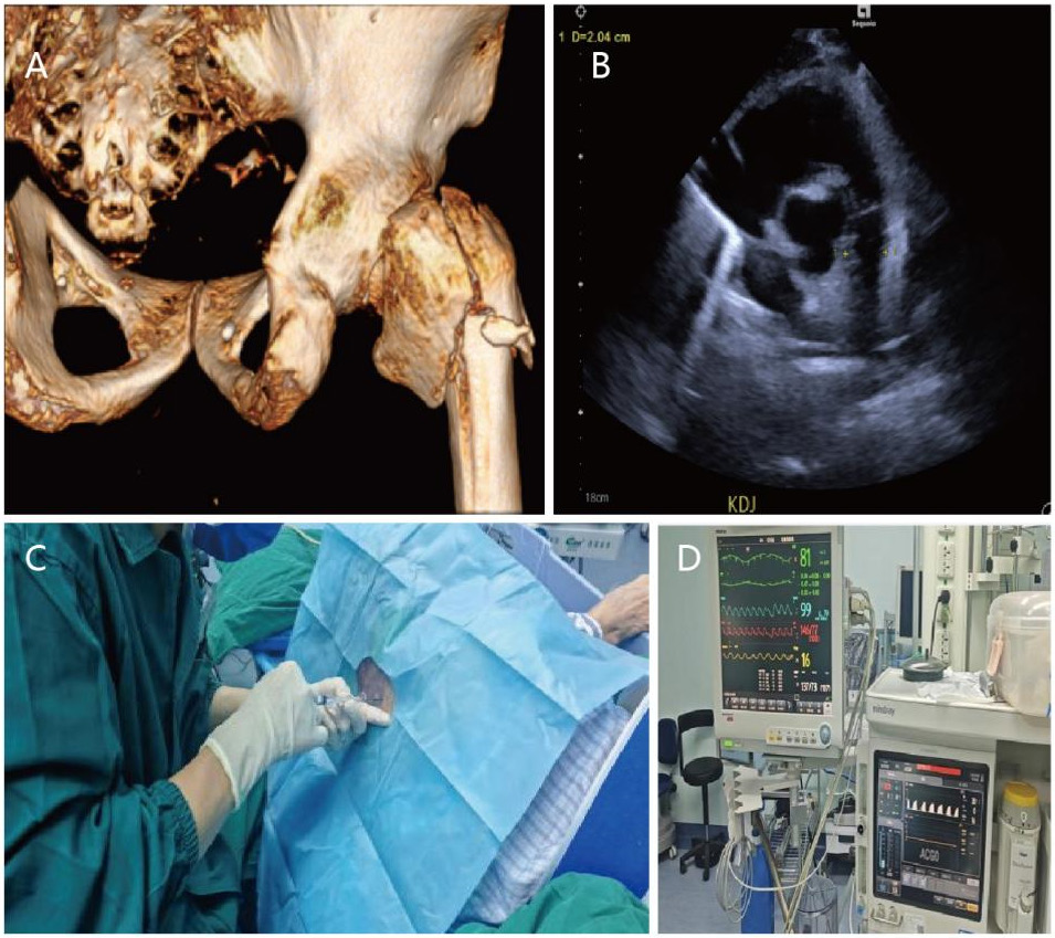
ventricular systolic function, and pulmonary artery pressure; (C) Ultrasound-assisted CSEA; (D) Intraoperative monitoring showing heart rate 81 beats/min, oxygen saturation 99%, and IBP 146/77 mmHg. CT, computed tomography; TTE, transthoracic echocardiography; CSEA, combined spinal–epidural anesthesia; IBP, invasive arterial blood pressure.
Anesthetic challenges: Maintaining appropriate preload, protecting right ventricular function, avoiding increases in pulmonary vascular resistance, and preserving systemic vascular resistance.
Management: Ultrasound-assisted CSEA was performed to achieve a T10 block level. IBP, CVP, and end-tidal CO2 were monitored. A 300 mL crystalloid preload was given. Dexmedetomidine (20 μg/h) provided sedation; norepinephrine (0.05–0.1 μg/kg/min) maintained blood pressure; milrinone (0.25–1.0 μg/kg/min) supported right ventricular function and reduced pulmonary vascular resistance.
Ultrasound application: Preoperative FICB; ultrasound-guided vascular access and neuraxial anesthesia; continuous intraoperative TTE monitoring of right ventricular size, interventricular septal motion, and tricuspid regurgitation to guide vasoactive and fluid therapy.
Outcome: Stable right ventricular function and hemodynamics were achieved, and the patient’s recovery in the ward was uneventful.
2.4 Case 4: Acute exacerbation of chronic heart failure—open reduction and internal fixation of left femoral fracture
A 97-year-old woman with a left intertrochanteric femoral fracture, congestive heart failure (New York Heart Association class III), coronary artery disease, and a history of treatment for chronic heart failure presented with dyspnea and bilateral pulmonary crackles on admission. NT-proBNP exceeded 30,000 pg/mL and troponin I was 0.554 μg/L. Echocardiography showed biatrial enlargement (left atrium 47 mm; right atrium 42 mm), interventricular septal thickening (12 mm), moderate mitral regurgitation, severe tricuspid regurgitation, mild aortic regurgitation, and impaired left ventricular diastolic function. After one week of medical therapy, NT-proBNP decreased to 22,213 pg/mL, and troponin I increased to 0.606 ug/L. The relevant images are shown in Figure 4.
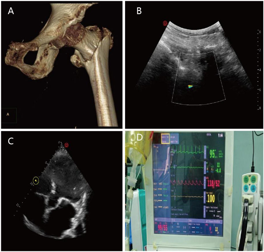
Anesthetic challenges: Extremely high surgical risk in the setting of acute heart failure, requiring optimization of preload, reduction of afterload, enhancement of myocardial contractility, and maintenance of effective cardiac output.
Management: The patient received 7 days of intensive care optimization (diuretics, vasodilators, low-dose dobutamine). After improvement, ultrasound-guided pericapsular nerve group block, vascular access, and low-dose CSEA (T10 level) were performed. IBP, CVP, and stroke volume variation guided fluid therapy. Norepinephrine (0.05–0.1 μg/kg/min) maintained blood pressure. Standard heart failure therapy was continued postoperatively.
Ultrasound application: Ultrasound-guided vascular access and nerve blocks; ultrasound-guided neuraxial anesthesia; TTE assessment of cardiac function and volume status.
Outcome: Intraoperative hemodynamics was stable. The patient was admitted to the Coronary Care Unit postoperatively and transferred to the general ward after one day. Cardiac function improved compared with before, and the patient was discharged successfully.
The clinical characteristics of the four cases are shown in Table 1.
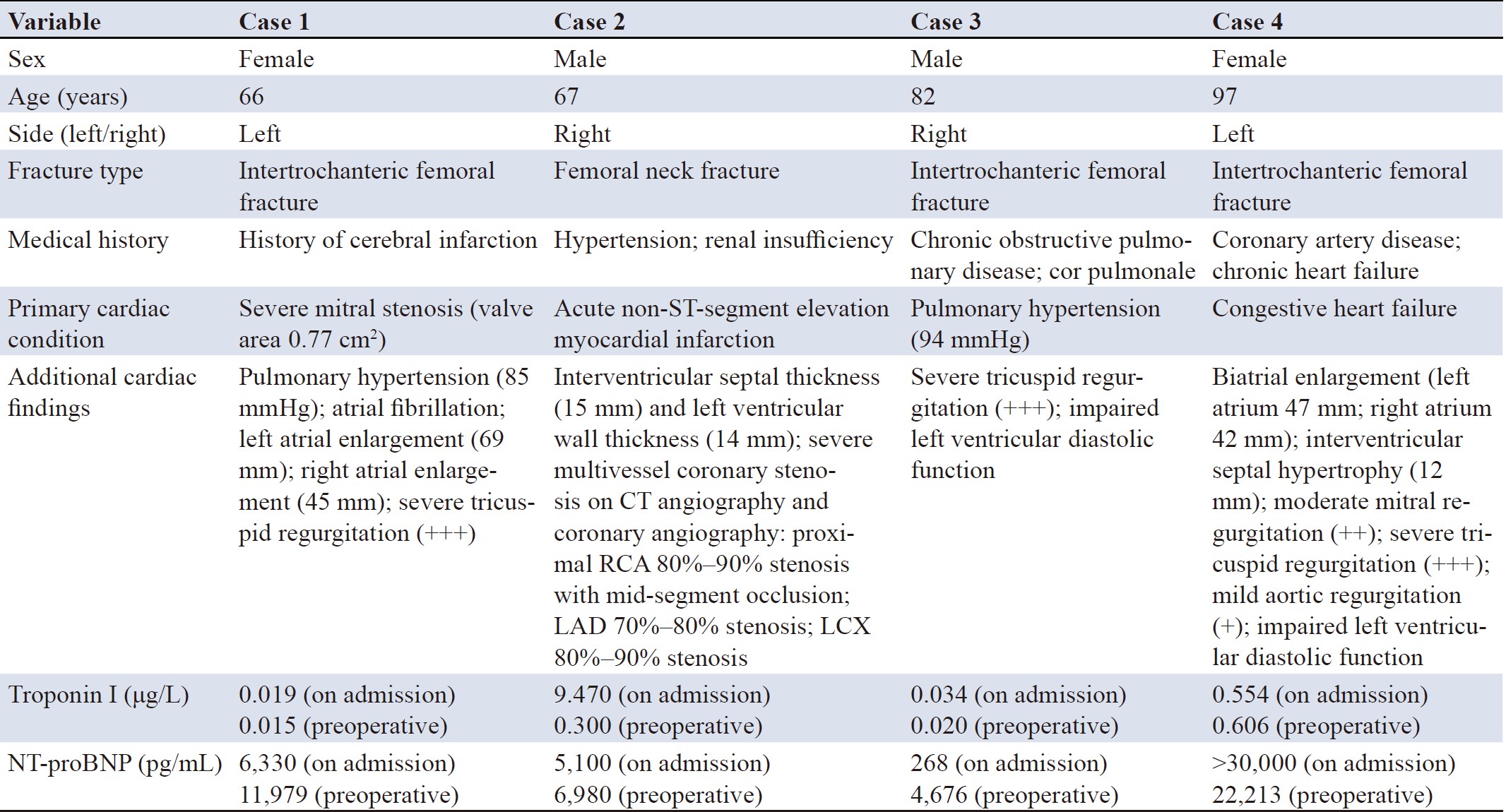
Note: CT, computed tomography; NT-proBNP, N‑terminal pro‑brain natriuretic peptide; RCA, right coronary artery; LAD, left anterior descending artery; LCX, left circumflex artery.
The successful anesthetic management of these four elderly hip fracture patients with severe cardiac disease highlights a paradigm shift from traditional experience-based practice to imaging- and function-guided precision anesthesia.
3.1 Core value and future trends of ultrasound
Perioperative ultrasound serves dual roles: Guiding regional anesthesia (e.g., FICB, neuraxial techniques), enabling the use of minimal effective doses while optimizing block quality and reducing sympathetic blockade and systemic drug effects; Enabling dynamic TTE monitoring, allowing direct visualization of cardiac structure and function, shifting from pressure-based to image-based assessment [2,3,8]. This is particularly critical in severe valvular disease, ventricular dysfunction, and complex heart failure, providing a direct basis for individualized management [1].
Future developments include portable intelligent devices (artificial intelligence-assisted measurements), full-process integration (pre-, intra-, and postoperative), integration with noninvasive hemodynamic monitoring, and remote ultrasound consultation to improve safety in resource-limited settings.
3.2 Pathophysiology-oriented hemodynamic management
Management strategies targeted disease-specific physiological priorities:
Mitral stenosis (Case 1): Heart rate control and maintenance of adequate preload to ensure diastolic filling [9].
Coronary artery disease (Case 2): Maintenance of myocardial oxygen supply–demand balance; strict avoidance of tachycardia and hemodynamic fluctuations [1].
Pulmonary hypertension (Case 3): Right heart–centered strategy emphasizing reduced pulmonary resistance, maintained perfusion, and enhanced right ventricular function [10,11].
Heart failure (Case 4): A continuum of optimization from preoperative stabilization to intraoperative support and postoperative analgesia to reduce cardiac workload and preserve pump function [12,13].
3.3 Cardioprotective role of multimodal analgesia
Multimodal analgesia based on nerve blocks combined with ultrasound-guided neuraxial anesthesia effectively interrupts the “pain–stress–cardiac load” cycle, reduces the need for general anesthetics, and provides clear cardioprotective benefits in patients with impaired cardiac function [8].
3.4 Limitations and future directions
Limitations include small sample size, lack of control group, non-standardized ultrasound monitoring, absence of long-term cardiac follow-up, and no comparison between regional anesthesia techniques.
Future directions include multicenter validation studies, artificial intelligence-assisted ultrasound decision-making models, closed-loop management guided by portable ultrasound, and health economic evaluations.
For elderly hip fracture patients with severe cardiac disease, anesthetic safety depends on: (1) ultrasound-enabled precision (from experience-based to visualization-guided practice); (2) a full-process strategy (preoperative optimization, intraoperative regulation, postoperative analgesia); (3) efficient multidisciplinary collaboration. Anesthesiologists should actively integrate ultrasound into perioperative care to reduce risk in this vulnerable population. Future advances in intelligent technologies and high-quality research are needed to further optimize management strategies.
Author contributions
Liangqing Lin conceived and designed the study, formulated the overall anesthesia strategy and major clinical decisions, screened the cases, and integrated the clinical data. Qinghua Wu performed and interpreted the perioperative ultrasound examinations, including assessments of cardiac function and volume status, and drafted the Methods and Results sections related to ultrasound techniques. Pinhui Ke and Chunlan Lin were primarily responsible for anesthetic management and intraoperative monitoring, collected perioperative clinical data from the four patients, including vital signs, medication records, and complications, and participated in data organization and preparation of the initial manuscript draft. Shuantong Lin assisted with the literature review and drafted the sections related to cardiac management strategies in the Discussion. Yaohua Yu reviewed the safety of the anesthesia protocols, provided additional clinical expertise regarding the application of ultrasound techniques, critically revised the discussion on anesthetic risks in patients with severe cardiac disease, and performed the final review and approval of the manuscript. All authors read and approved the final manuscript.
Funding
This study was supported by the Putian Science and Technology Program 2025 (2025NJYL026).
Data availability
The de-identified individual participant data are available from the corresponding author (Yaohua Yu, yyh.8@163.com) upon reasonable request.
Ethics approval and consent to participate
This study was approved by the Ethics Committee of The First Hospital of Putian (Approval No. 2026-059). Written informed consent was obtained from all participants or their legal representatives.
Consent for publication
Written informed consent for publication was obtained from all patients or their legal representatives.
Competing interests
The authors declare no conflicts of interest related to this study.
Acknowledgements
None.
[1] Trauzeddel RF, Ertmer M, Nordine M, Groesdonk HV, Michels G, Pfister R, et al. Perioperative echocardiography-guided hemodynamic therapy in high-risk patients: A practical expert approach of hemodynamically focused echocardiography. J Clin Monit Comput. 2021 Apr;35(2):229-243. https://doi.org/10.1007/s10877-020-00534-7
[2] Thompson A, Fleischmann KE, Smilowitz NR, de Las Fuentes L, Mukherjee D, Aggarwal NR, et al. 2024 AHA/ACC/ACS/ASNC/HRS/SCA/SCCT/SCMR/SVM guideline for Perioperative cardiovascular management for noncardiac surgery: A report of the American College of Cardiology/American Heart Association Joint Committee on Clinical Practice Guidelines. Circulation. 2024 Nov 5;150(19):e351-e442. https://doi.org/10.1161/CIR.0000000000001285
[3] Duceppe E, Parlow J, MacDonald P, Lyons K, McMullen M, Srinathan S, et al. Canadian cardiovascular society guidelines on perioperative cardiac risk assessment and management for patients who undergo noncardiac surgery. Can J Cardiol. 2017 Jan;33(1):17-32. https://doi.org/10.1016/j.cjca.2016.09.008
[4] Neuman MD, Ellenberg SS, Sieber FE, Magaziner JS, Feng R, Carson JL, et al. Regional versus general anesthesia for promoting independence after hip fracture (REGAIN): Protocol for a pragmatic, international multicentre trial. BMJ Open. 2016 Nov 15;6(11):e013473. https://doi.org/10.1136/bmjopen-2016-013473
[5] Vail EA, Feng R, Sieber F, Carson JL, Ellenberg SS, Magaziner J, et al. Long-term outcomes with spinal versus general anesthesia for hip fracture surgery: A randomized trial. Anesthesiology. 2024 Mar 1;140(3):375-386. https://doi.org/10.1097/ALN.0000000000004807
[6] Lenk T, Whittle J, Miller TE, Williams DGA, Bronshteyn YS. Focused cardiac ultrasound in preoperative assessment: The perioperative provider’s new stethoscope? Perioper Med (Lond). 2019 Nov 22;8:16. https://doi.org/10.1186/s13741-019-0129-8
[7] Canty DJ, Heiberg J, Yang Y, Royse AG, Margale S, Nanjappa N, et al. Pilot multi-centre randomised trial of the impact of pre-operative focused cardiac ultrasound on mortality and morbidity in patients having surgery for femoral neck fractures (ECHONOF-2 pilot). Anaesthesia. 2018 Apr;73(4):428-437. https://doi.org/10.1111/anae.14130
[8] Exsteen OW, Svendsen CN, Rothe C, Lange KHW, Lundstrøm LH. Ultrasound-guided peripheral nerve blocks for preoperative pain management in hip fractures: A systematic review. BMC Anesthesiol. 2022 Jun 21;22(1):192. https://doi.org/10.1186/s12871-022-01720-7
[9] Galusko V, Ionescu A, Edwards A, Sekar B, Wong K, Patel K, et al. Management of mitral stenosis: A systematic review of clinical practice guidelines and recommendations. Eur Heart J Qual Care Clin Outcomes. 2022 Sep 5;8(6):602-618. https://doi.org/10.1093/ehjqcco/qcab083
[10] Haddad F, Hunt SA, Rosenthal DN, Murphy DJ. Right ventricular function in cardiovascular disease, part I: Anatomy, physiology, aging, and functional assessment of the right ventricle. Circulation. 2008 Mar 18;117(11):1436-1448. https://doi.org/10.1161/CIRCULATIONAHA.107.653576
[11] Ramakrishna H. Pulmonary hypertension in the perioperative period-focus on current and emerging therapies. Recent Pat Cardiovasc Drug Discov. 2014;9(1):38-50. https://doi.org/10.2174/1574890109666141024150509
[12] Metra M, Adamo M, Tomasoni D, Mebazaa A, Bayes-Genis A, Abdelhamid M, et al. Pre-discharge and early post-discharge management of patients hospitalized for acute heart failure: A scientific statement by the Heart Failure Association of the ESC. Eur J Heart Fail. 2023 Jul;25(7):1115-1131. https://doi.org/10.1002/ejhf.2888
[13] Metra M, Tomasoni D, Adamo M, Bayes-Genis A, Filippatos G, Abdelhamid M, et al. Worsening of chronic heart failure: Definition, epidemiology, management and prevention. A clinical consensus statement by the Heart Failure Association of the European Society of Cardiology. Eur J Heart Fail. 2023 Jun;25(6):776-791. https://doi.org/10.1002/ejhf.2874
ISSN: 2957-5443
Volume 4, Issue 3
September 2026

 Log In

 On This Page
 CITE
CITE