 Log In
Search




 Open Access
 h_b_cui@163.comOpen Accessh_b_cui@163.com
Combat trauma hemostasis techniques are crucial for modern military medicine in addressing the challenges posed by high-energy destructive weapons. As reported, the incidence of vascular injuries during the Vietnam War was approximately 2%, while that during the Iraq War ranged from 4.4% to 8.2%. Meanwhile, massive hemorrhage caused by various types of vascular injuries is the primary factor leading to acute death among potentially survivable casualties during wartime. This paper provides a detailed exploration of the research progress in combat trauma hemostasis, with a focus on analyzing vascular injuries and uncontrolled bleeding caused by high-energy destructive weapons in modern warfare. Starting from the pathophysiological characteristics of combat trauma, it introduces the four-level priority treatment system established by North Atlantic Treaty Organization and Committee on Tactical Combat Casualty Care, and emphasizes the importance of the “Golden 1-Hour” principle in improving casualty survival rates. The physiological mechanisms of blood coagulation are outlined, followed by an in-depth analysis of both traditional and novel hemostatic techniques, including tourniquets, hemostatic dressings, and auxiliary hemostatic materials. Finally, the challenges faced by combat trauma hemostasis technologies are discussed, along with future development directions.
In the injury spectrum of modern warfare, explosive fragments and gunshot wounds account for over 75% of battlefield casualties, where uncontrollable hemorrhage is the primary pathological factor leading to on-site fatalities among casualties [1]. Notably, with the continuous iteration and upgrading of high-energy destructive weapons, the incidence of vascular injuries has shown a significant upward trend. During the Vietnam War, the occurrence rate of vascular trauma was merely 2% [2, 3]. During the Iraq War, this figure rose to 4.4%-8.2% [4, 5]. More recent analyses from conflicts in Afghanistan and Syria have suggested that vascular injury rates can reach 9-12% in conventional combat scenarios [6]. Research on combat trauma treatment has confirmed that fatal massive hemorrhages caused by various types of vascular injuries are the primary cause of acute death among potentially survivable battlefield casualties, with their pathological significance far exceeding that of other trauma types [7].
The evolution of modern warfare has further intensified the complexity of combat casualty care. Under the three-dimensional combat mode supported by sixth-generation communication technology and the application of new equipment such as high-explosive munitions and thermobaric weapons, casualties commonly exhibit the “four highs” characteristics—namely, a high proportion of kinetic energy injuries (>68%), a high incidence of combined injuries (41%), a high degree of tissue destruction, and a high risk of secondary hemorrhage [7, 8]. Taking a 70 kg healthy adult male as an example, the circulating blood volume accounts for approximately 7%-8% of body weight (about 4,900 ml). When acute blood loss exceeds 30% of the total blood volume (i.e., 1,470 ml), it can lead to decompensated shock. Among battlefield fatalities, 56% are directly attributed to uncontrolled bleeding, with this proportion rising to as high as 83% in preventable prehospital deaths [9]. Clinical big data analysis reveals that among fatal complications such as multiple organ dysfunction syndrome and disseminated intravascular coagulation following combat trauma, 61.2% of cases involve uncontrolled progressive hemorrhage, with over 18% of these deaths potentially preventable through early and effective hemostatic intervention [10].
Given the unique pathophysiological characteristics of combat injuries, domestic vascular surgery experts, based on the analysis of outcomes from the management of vascular combat trauma during years of military operations and training, propose that the treatment of vascular combat trauma should adhere to the “Three Rapid” principles: “rapid diagnosis, rapid hemostasis, and rapid revascularization” [3]. The Committee on Tactical Combat Casualty Care of North Atlantic Treaty Organization has established a four-tier priority management system: Immediate, Delayed, Minimal, and Expectant [11]. These principles and systems operate within a framework constrained by three major contradictions in combat casualty care: 1) the contradiction between the probability of mass casualties and limited medical resources; 2) the contradiction between complex environments (such as high-altitude hypoxia and high humidity in maritime areas) and the effectiveness of hemostatic materials; 3) the contradiction between multi-echelon evacuation (including three levels of care: tactical zone, operational zone, and strategic zone) and the continuity of hemostatic measures. Under this challenging framework, the “Golden Hour Rule” emphasizes that for every 10-minute reduction in the time window from injury to definitive hemostasis, the survival rate of casualties increases by 7.3% [12]. This stringent timeliness requirement arises directly from the need to overcome these contradictions. Therefore, the development of a dual-mode hemostatic technology system that combines “rapid response in frontline first aid” with “compatibility and scalability for rear surgical procedures” has become a strategic priority in 21st-century military medical research.
Hemostasis on the battlefield involves a combination of mechanical occlusion, biochemical coagulation, and thermal method, all ultimately relying on the body’s intrinsic coagulation cascade. Vascular occlusion primarily relies on physical methods, with compression being the main hemostatic approach at body junctions [13]. For instance, after bleeding occurs, rapid hemostasis is achieved by using fingers to compress the superficial area of an artery overlying a bony surface (proximal end), thereby blocking blood flow. This method is simple to perform and suitable for hemostasis in areas such as the head, neck, shoulders, and limbs, but it is difficult to maintain for extended periods. Other tools like tourniquets and hemostatic forceps can also be employed to swiftly cut off blood flow at the bleeding site for rapid hemostasis. Blood coagulation involves multiple physiological regulatory levels, including key steps such as platelet activation, the coagulation cascade, and fibrin formation. When blood vessels are damaged, initial vasoconstriction occurs, slowing blood flow to reduce blood loss. Platelets at the injury site become activated, adhere to the damaged area, and aggregate to form an initial platelet plug—a process known as primary hemostasis, which provides only temporary hemostatic effects [14]. The secondary hemostasis is formed by the combination of fibrin polymers and platelet plugs, creating a firm and durable thrombus that occludes the wound for an extended period to achieve hemostasis.
The entire coagulation cascade consists of two main pathways: the intrinsic pathway (contact activation) and the extrinsic pathway (tissue factor). Both pathways initiate the common pathway by activating Factor X. Activated thrombin converts soluble fibrin into insoluble fibrin, which then binds to platelet plugs to form a stable thrombus, effectively halting blood flow [15].
The hemostatic principle of electrocautery in surgical procedures is primarily based on thermal effects, achieving hemostasis by heating tissues. During surgery, the electrocautery device delivers high-frequency electrical currents or laser waves, which act on the target tissue and cause a rapid localized temperature increase. Specifically, the high temperature evaporates water within tissue cells, leading to cellular desiccation and coagulation as moisture is lost. This process not only directly disrupts cellular structures but also induces contraction of surrounding vascular smooth muscle, reducing blood flow and enabling rapid hemostasis. Simultaneously, the thermal effect activates the coagulation cascade, promoting platelet aggregation and activation to form thrombi, further sealing the wound. Crucially, this thermal coagulation method offers high precision, effectively controlling bleeding without damaging adjacent healthy tissues. The use of electrocautery in surgery significantly reduces intraoperative blood loss, shortens ope-rative time, and enhances procedural safety and efficacy. Consequently, electrocautery is widely employed in various surgical interventions, such as tumor resection and visceral organ surgeries, to ensure smooth procedures and favorable postoperative recovery outcomes.
Additionally, advancements in temperature control technology for surgical electrocautery continue to improve, minimizing excessive thermal damage during hemostasis and further enhancing safety and effectiveness. For instance, modern electrocautery devices may incorporate intelligent temperature monitoring systems that track real-time temperature fluctuations and automatically adjust power output to maintain optimal hemostatic conditions. Such high-tech applications not only improve hemostatic performance but also reduce complication risks, thereby elevating the overall success rate of surgical procedures.
Traditional hemostatic methods for combat trauma primarily include direct manual pressure, mechanical compression devices like tourniquets, and passive hemostatic dressings. Digital pressure hemostasis is a method where, after bleeding occurs, fingers are used to compress the relatively superficial portion of an artery above a bony surface (proximal end) to block blood flow and achieve rapid hemostasis. This technique is operationally simple and suitable for hemostasis in areas such as the head, neck, shoulders, and limbs, but it is difficult to maintain for extended periods [16]. Traditional hemostatic equipment such as hemostatic gauze and hemostatic cotton (subtypes of hemostatic dressings) is primarily used for packing and compression, with limited hemostatic effects and lacking other hemostatic mechanisms, thus making them difficult to meet the hemostatic demands of traumatic massive hemorrhage.
Among mechanical devices, the tourniquet, as a cornerstone technique for controlling massive hemorrhage in combat trauma, has a history of application dating back to the Hippocratic era of ancient Greece. Modern war medicine data indicate that standardized use of tourniquets can reduce mortality from extremity arterial injuries from 23.8% to 4.1%, with individual soldier compliance rates exceeding 92% [17]. The core mechanism lies in completely blocking distal arterial blood flow through mechanical compression (pressure threshold >250 mmHg), creating an “ischemia-hemostasis time window”. The Tactical Field Assessment, as a standardized procedure for hemorrhage identification, requires systematic visual and tactile examination of 12 key anatomical points such as the neck, axilla, and groin to promptly identify life-threatening bleeding signs. These include traumatic limb amputation, arterial pulsatile bleeding, blood saturation of clothing (>500 ml/m²), and compensated shock manifestations (altered mental status, systolic blood pressure <90 mmHg). This assessment system incorporates tourniquet application timeliness into the golden treatment standard: completing"High-and-Tight" binding with-in 1 minute of contact with the casualty achieves optimal outcomes, with a 7.3% survival rate decrease for every 10-minute delay [12].
Based on the anatomical characteristics of the injury, traditional hemostatic devices are classified into four major categories: 1) extremity tourniquets: these devices employ a dual-loop windlass design (e.g., Combat Application Tourniquet Generation 7), achieving self-locking pressure via nylon self-adhesive straps, and are suitable for controlling proximal humerus/femur hemorrhage; 2) junctional tourniquets: this category includes devices such as the SAM® Junctional Tourniquet (SJT), which utilizes rigid pressure plates to apply trans-soft-tissue compression on the iliac artery; 3) hemostatic dressings: these comprise materials like chitosan gauze (Celox®) and kaolin dressing (QuikClot®), which achieve wound hemostasis through charge adsorption or coagulation factor activation; 4) auxiliary hemostatic materials: these are substances such as hemostatic powder (WoundStat®) and fibrin glue (Tisseel®), used for supplementary hemostasis in specialized wound types [18].
Taking extremity tourniquets as an example, their technological evolution has undergone three generations of innovation: 1) the first generation (simple cloth bandage): it was widely used during the Vietnam War, with a secondary injury rate as high as 34%; 2) the second generation (elastic rubber tourniquet): it was deployed during the Gulf War, increasing hemostasis success rate to 79%; 3) the third generation (composite self-locking bandage): it was popularized after the Iraq War, utilizing polyester/nylon composite webbing and ratchet buckle, achieving a hemostasis success rate >96% and limb salvage rate of 89% [19].
However, traditional hemostatic techniques still exhibit significant limitations: 1) the time-efficacy-injury paradox: tourniquet use exceeding 2 hours can lead to nerve damage (incidence rate of 19.7%) and rhabdomyolysis; 2) anatomical blind zones: this refers to the inefficient control of subclavian artery and pelvic hemorrhage, with a success rate below 65%; 3) environmental sensitivity: dressing adhesion decreases by 37% in high-humidity environments, while low temperatures reduce thrombin activity by 52% [20].
The rapid development of materials science has driven the innovation of hemostatic dressings, progressively replacing traditional hemostatic powders with advanced forms like hemostatic sponges and gels. Hemostatic materials can be roughly divided into three categories: polysaccharides, inorganic minerals, and proteins, each represented by various advanced formulations [21]. Polysaccharide-based materials, particularly chitosan, are widely explored. Chitosan is a natural polysaccharide extracted from shrimp and crab shells, possessing biocompatibility, biodegradability, non-toxicity, and antibacterial properties [22]. These characteristics make it an ideal absorbable hemostatic material, capable of reducing immune responses, minimizing complications, avoiding infections caused by long-term retention, and promoting wound healing. The XStat hemostatic injector is a 3 cm diameter polycarbonate syringe containing a hemostatic sponge. The XStat injectable hemostatic device achieves hemostasis through the rapid expansion mechanism of chitosan-coated cellulose sponges (Figure 1), providing a novel solution for addressing deep non-compressible hemorrhage. This device developed by the U.S. military injects 92-100 chitosan-coated cellulose sponges (9 mm diameter × 4.5 mm height) into the wound cavity, which expands 12-fold within 12 seconds to form a three-dimensional pressure matrix. The preclinical trial of XStat was conducted using the U.S. military standard porcine femoral artery injury model (creating a 6 mm diameter hole in the porcine femoral artery), comparing its hemostatic effect with that of standard hemostatic dressings [23]. The results showed: In terms of time required to complete hemostasis, the XStat 30 group [(1.1±0.3) min] was significantly shorter than the conventional standard wound dressing group [(4.6±0.5) min]; regarding the amount of blood loss (measured from 45 seconds after creating the femoral artery hole until the end of the 6-hour observation period), the XStat group [(21.7±17.5) ml] had significantly less blood loss than the conventional standard wound dressing group [(28.3±18.1) ml] [6, 10]. In both groups, all trial pigs achieved 100% hemostasis and 100% survival. These results indicate that both XStat and standard hemostatic dressings have good hemostatic effects, but the former achieves hemostasis more rapidly.

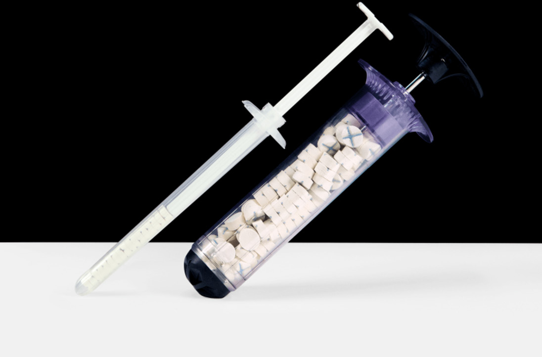
Figure 1. XStat hemostatic injector. The device consists of a syringe (for delivering sponges) and chitosan-coated cellulose sponges (core hemostatic component). The sponges expand rapidly after contact with blood to form a pressure matrix.
However, the cellulose matrix of this device is non-degradable, and delayed removal will increase the incidence of granuloma formation to 17%, while high humidity environments reduce its expansion rate by 29% [19, 20].
Inorganic minerals, such as kaolin dressings, accelerate coagulation through the factor XII contact activation pathway but are limited by increased risk of failure in patients with trauma-induced coagulopathy [24]. This technical difference highlights the need to strike a balance in the development of hemostatic materials between the selectivity of coagulation mechanisms (factor-dependent/independent), material degradability, and environmental adaptability. The physical hemostatic function of kaolin is similar to that of zeolite-based materials, as both belong to aluminosilicate minerals capable of absorbing moisture from blood, concentrating clotting factors, and rapidly initiating the coagulation system [25]. Since kaolin primarily functions by activating the endogenous coagulation pathway, its hemostatic effect significantly diminishes when massive blood loss leads to substantial depletion of coagulation factors and secondary coagulation dysfunction [26]. The self-developed emergency chitosan hemostatic sponge, through positive charge-mediated erythrocyte aggregation and combined with the super absorbency (water absorption ratio ≥40 g/g) of polyacrylic resin dressings, can synergistically control deep laceration bleeding [27]. The rapid hemostatic powder “Blood Shield” uses modified zeolite as its core component. Through a dual mechanism of calcium ion release and wound charge adsorption, it achieves control of arterial and venous bleeding within 30 seconds (hemostatic efficacy rate >94%). It has been standardized as essential individual first-aid equipment [28].
Protein-based materials like fibrin glue (Tisseel®) are also used (See Table 1). Furthermore, composite materials combining different mechanisms are promising. The military transformation potential of traditional Chinese medicine hemostatic materials is gradually becoming evident. For instance, based on research guided by traditional medical theories, a hemostatic agent composed of Bletilla striata and zeolite was developed by optimizing the components. The formulation combines Bletilla striata lyophilized powder (the non-polysaccharide fraction of the 80% ethanol extract of Bletilla striata) with nano-zeolite at a 3:7 ratio, reducing coagulation time to 1.8±0.3 minutes. Additionally, coating the zeolite with Bletilla striata polysaccharides lowered the peak heat generation (ΔT decreased from 42 °C to 31 °C), resulting in a reduction of burn incidence from 28% to 6% [31]. Meanwhile, the 37-Chitosan synergistic system demonstrates advantages in multi-target regulation. Panax notoginseng saponins (R1+Rg1≥8%) can activate platelet GPⅡb/Ⅲa receptors, and synergize with chitosan to promote fibrin cross-linking, thereby achieving significant effects in combat wound repair. Animal experimental results indicate that the wound healing rate is 37% faster than that of traditional dressings [30, 32].
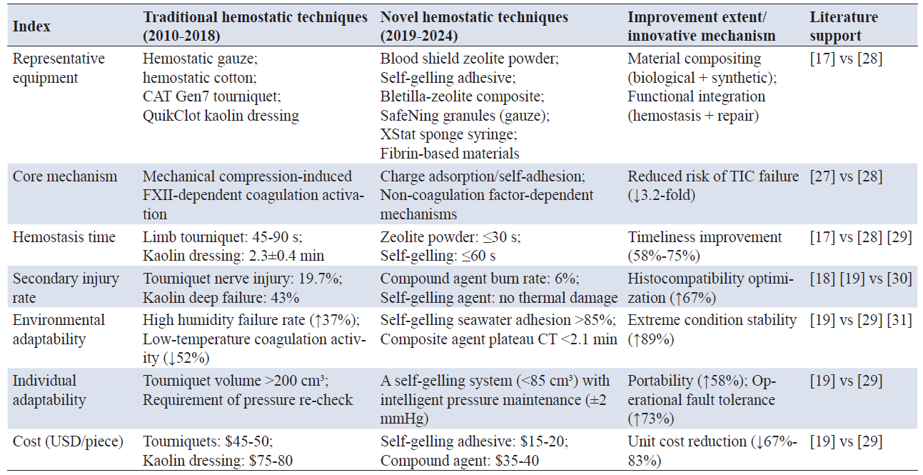
Note: CAT Gen7, Combat Application Tourniquet, Generation 7; FXII, Coagulation Factor XII; TIC, Trauma-Induced Coagulopathy; CT, Coagulation Time; USD, United States Dollar.
Our military has achieved a series of breakthroughs in the field of hemostatic material development, establishing a multidimensional technical system that encompasses physical occlusion, biochemical activation, and composite mechanisms. The independently developed emergency chitosan hemostatic sponge induces red blood cell aggregation through positive charge mediation, while the superabsorbent polyacrylic resin dressing (water absorption ratio ≥40 g/g) synergistically controls deep cavity bleeding [27].
Another notable advancement is the ultrafast self-gelling/wet-state powdered adhesive developed by the research team of Bian Liming at The Chinese University of Hong Kong. It has broken through the limitations of traditional formulations, exhibiting superior wet-state adhesion that achieves a bonding strength exceeding 50 kPa within one minute on hydrated wound surfaces. Simultaneously, its excellent topological adaptability enables effective filling of irregular wound cavities, with a compatibility rate exceeding 92%. Additionally, this material demonstrates significant cost advantages, with the production cost per unit dose being only 17% of commercially available hemostatic agents [29]. After undergoing tactical environment simulation tests, the material maintained stable hemostatic efficacy under seawater immersion and high-altitude low-pressure conditions.
This evolution in technical approaches highlights the critical need for balancing selectivity in coagulation mechanisms (factor-dependent vs. independent), material degradability, and environmental adaptability in the development of next-generation hemostatic materials. Table 1 summarizes the advantages and disadvantages of different generations of hemostatic materials for combat trauma.
Currently, the cuff-inflated pneumatic tourniquet is considered the optimal choice with high hemostatic efficiency, optimal patient comfort, and the lowest complication rates. It consists of an inflatable balloon, a pressure-release valve, and a self-adhesive inflatable cuff. Its primary drawback is skin injury. Throughout the entire application process of the tourniquet, any minor issue leading to improper use can result in skin damage, such as redness, bruising, or necrosis. Additionally, the device’s susceptibility to damage, low reusability, and the difficulty for non-medical professionals to accurately control cuff pressure have prevented its widespread frontline deployment. During the research and development process, if intelligent elements could be integrated, such as enabling a "one-touch" automatic pressurization function and utilizing ultrasound or tactile sensors to automatically assess arterial compression, it would not only ensure distal blood supply while achieving hemostasis but also automatically calculate hemostasis duration and prompt timely release [33]. In this way, military medical personnel can allocate more time to rescuing other casualties.
The intelligent design has endowed this device with immense imaginative potential. In the future, inflatable hemostatic devices could be pre-assembled in junctional areas such as the neck, axillae, and groin of combat uniforms or protective gear, without interfering with soldiers' tactical movements during operations. In the event of major limb hemorrhage, a "high and tight" hemostatic effect can be achieved by triggering a switch; for junctional zone bleeding, effective compression of the bleeding area can be realized. Beyond mechanical function, research suggests integrating casualty identification systems and BeiDou positioning systems onto new-generation tourniquets [34]. The integration of intelligent elements will enable tourniquets to play an even more crucial role in modern warfare.
Traditional emergency hemostatic methods often require medical personnel to dress wounds in hazardous environments, undoubtedly increasing the risks of on-site rescue operations and even potentially endangering the safety of the rescuers themselves. Consequently, employing unmanned rescue equipment such as robotic arms to replace manual intervention holds critical significance for saving lives, alleviating the workload pressure on medical staff, and mitigating personnel shortages in healthcare. Several other widely used hemostatic devices and methods currently in practice are introduced below.
The combat-ready hemostatic clamp (Figure 2) weighs approximately 700 g and consists of a C-shaped steel support frame and a plastic pressure disk, serving as a clamp-type hemostatic device. This apparatus can directly apply pressure to wounds or junctional areas, compressing blood vessels to achieve effective hemostasis, making it particularly suitable for amputated limb hemorrhage control [35]. The combat ready clamp is the first junctional hemorrhage control device issued to foreign military forces, demonstrating relatively ideal hemostatic efficacy. However, it requires considerable time for assembly and is prone to detachment during transport [16].
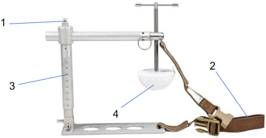
Figure 2. Combat ready clamp. 1, T-shaped threaded adjustable rod; 2, Fixing belt; 3, C-shaped steel bracket; 4, Plastic compressor.
The iTClamp hemostatic clamp (Figure 3) is manufactured by the Canadian company iTraumaCare. Its appearance resembles that of a common clip, weighing 37.1 grams, and is suitable for hemostasis in limbs, armpits, groin, and head/neck regions. Compared to traditional instruments, this hemostatic device employs an innovative approach by approximating wound edges to form an internal hematoma, thereby compressing damaged blood vessels and effectively controlling bleeding [36]. Unlike conventional hemostatic methods, the iTClamp design not only simplifies the operational procedure but also enhances comfort at the wound site, preventing excessive pain caused by traditional approaches for patients. In 2019, the updated U.S. Tactical Combat Casualty Care guidelines still recommended iTClamp for controlling major hemorrhage [34]. The iTClamp approximates wound edges through four pairs of opposing needles, utilizing the hydrostatic pressure generated by the hematoma to further control bleeding [16]. This hemostatic method not only does not increase pain at the wound site, but it also does not cause tissue necrosis even with prolonged use. Shang Z et al. employed a robotic arm to manage limb hemorrhage in casualties, where under the constraint of an external rigid structure, effective hemostasis was achieved through radial deformation of the airbag to compress the limb [37].
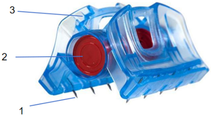
Figure 3. iTClamp hemostatic clamp. 1, Four pairs of opposing needle set; 2, Ergonomic clip body; 3, Anti-slip contact surface.
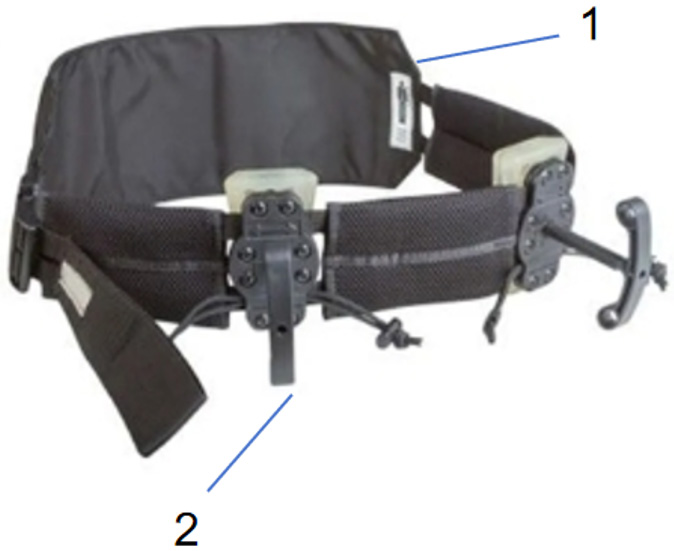
Figure 4. Junctional emergency treatment tool. 1, Nylon waistband; 2, Two T-shaped adjustable handles with pressure pads.
The SJT (Figure 5) is manufactured by the American company Sam Medical Products. It resembles the JETT in appearance, weighs 450 grams, and differs from the JETT in that the SJT is equipped with two inflatable cuffs. It can also be used for pelvic fractures, with a maximum usage duration of 4 hours [38]. The SJT was designed with ergonomic considerations, allowing it to fit snugly against the injured limb during use to ensure effective hemostasis. Its operation is simple, requiring medical personnel only to inflate the cuff using the accompanying air pump device to quickly achieve hemostasis. Additionally, the SJT is equipped with a pressure monitoring system that can track cuff pressure in real time, ensuring stable pressure during hemostasis and preventing tissue damage from excessive compression.
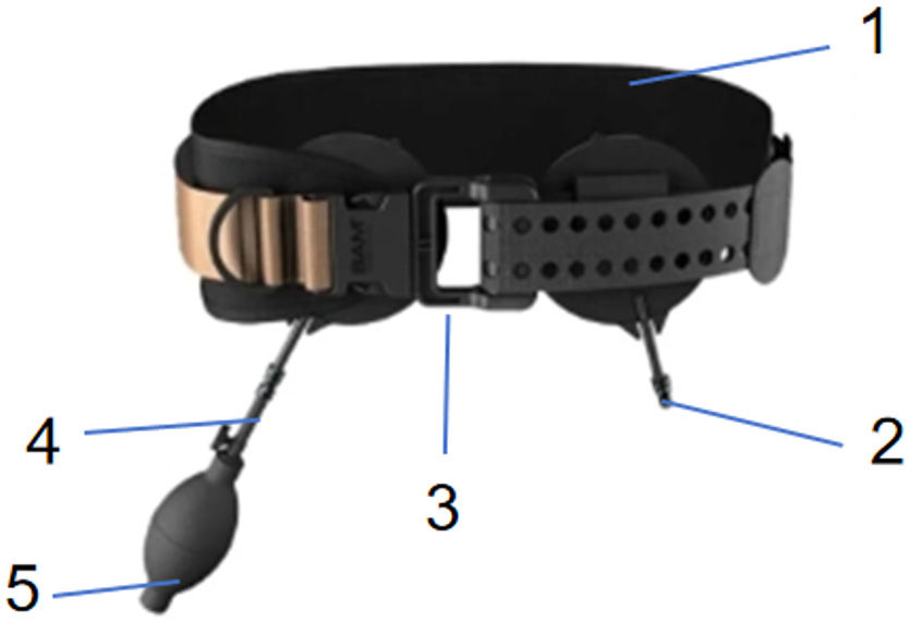
Figure 5. SAM® junction tourniquet. 1, Pressure pad expander; 2, Auxiliary Frenulum; 3, Pressure pad; 4, Pressure regulator; 5, Inflatable balloon.
High-frequency electrocoagulation technology is a minimally invasive technique that utilizes the heat generated by high-frequency current to coagulate tissues, thereby achieving therapeutic purposes such as cutting, hemostasis, and ablation. Due to its minimally invasive, safe, and effective characteristics, it has been increasingly widely applied in clinical practice. However, existing high-frequency electrocoagulation devices adopt a structure combining a main unit with a handpiece, which is bulky, accessory-intensive, and cumbersome to use. Common electrocoagulation handpieces also have a limited hemostatic area and low efficiency. Consequently, current high-frequency electrocoagulation hemostasis techniques cannot meet the demands of combat trauma care, such as large-area traumatic bleeding, mass casualty treatment, and time-sensitive emergency scenarios.
As a representative of our military’s new hemostatic equipment, the portable field high-frequency hemostatic device achieves rapid hemostasis through the thermal coagulation effect of high-frequency current (2 MHz). Its core innovation lies in the tactical adaptation of traditional high-frequency electrosurgical technology. The device employs a bipolar coagulation mode, where precision forceps electrodes with a spacing of ≤3 mm act precisely on the bleeding site, raising local tissue temperature to 80-100 °C within 10-15 seconds to induce vascular closure. Simultaneously, the depth of thermal damage is controlled within 1.2 mm, significantly reducing the risk of tissue necrosis. The total weight of the unit is <1.5 kg, equipped with a lithium-ion battery and direct current/alternating current conversion module, enabling continuous operation for 45 minutes in environments without a mains power supply, and adaptable to complex battlefield conditions such as high-altitude, humid, and hot climates. Clinical tests demonstrate an 88% control rate for deep non-compressible hemorrhage, making it particularly suitable for irregular wounds such as shrapnel embedding injuries [39]. However, the device still requires operation by professionals, and its cost is much higher than traditional tourniquets. In the future, the integration of impedance sensing and artificial intelligence positioning modules could further enhance individual soldier adaptability.
The core challenge faced by battlefield trauma hemostasis technology lies in striking a balance between efficiency, safety, and battlefield adaptability. Although traditional hemostatic methods such as tourniquets and kaolin dressings are widely used in combat settings, they struggle to overcome several critical limitations. For instance, mechanical compression may lead to nerve injuries, with an incidence rate as high as 19.7%. Meanwhile, the failure rate for deep wound hemostasis can reach 43%. Additionally, the performance degradation of these conventional methods in low-temperature or high-humidity environments cannot be overlooked. Research indicates that coagulation activity may decrease by up to 52%.
Although the novel high-frequency electrocoagulation technology can achieve rapid hemostasis through thermal effects, with the entire process taking only 8-12 seconds, it still faces challenges such as thermal damage control (thermal damage depth ≤1.5 mm), bulky equipment (weight exceeding 5 kg), and high costs (unit price approximately $1,200) [33]. Furthermore, extreme battlefield environments (such as high-altitude hypoxia or high-salt fog in maritime areas) impose even more stringent requirements on the stability of hemostatic materials. Current technologies have yet to fully meet the demands for mass casualty treatment across diverse scenarios and varying injury types.
Under the current circumstances, innovative concepts such as intelligence, integration, lightweight design, miniaturization, recyclability, high comfort, strong stability, and low cost will provide significant reference value for the development of novel hemostatic medical devices. The future trend of hemostatic technology may focus on the application of electromagnetic induction principles—for instance, utilizing non-contact energy transfer to achieve safe and efficient new hemostatic equipment. Such devices can achieve complete isolation between the human body and the power source, fundamentally eliminating risks that traditional hemostatic methods may pose to both patients and medical staff. In contrast, traditional surgical electrocautery relies on human impedance as part of the circuit, employing high-frequency, high-voltage currents. This not only increases the risk of electric shock and burns for patients but also exposes them to direct electric shock in case of equipment failure.
Specifically, the new equipment will adopt an innovative safety design aimed at ensuring hemostatic efficiency while improving operational safety. This not only reduces surgical risks but also enhances applicability in extreme battlefield environments, better meeting the demands of modern warfare for hemostatic technology. With continuous technological advancements, future hemostatic devices will evolve toward greater intelligence, portability, and efficiency. For example, the development of nanotechnology enables micro-robots to perform precise hemostatic operations within the human body. In the future, nanorobots may be utilized to repair damaged blood vessels and control bleeding or achieve localized drug release to promote wound healing and hemostasis. Progress in bioprinting technology makes it possible to print tissues with vascular systems, which holds significant implications for hemostatic treatment and tissue repair in combat-wounded patients, providing more reliable solutions for battlefield medical care.
The advancement of battlefield trauma hemostasis technology represents a pivotal component of modern military medicine for addressing the challenges posed by high-energy destructive weapons. From the mechanical compression of traditional tourniquets to the precise thermal effects of high-frequency electrocoagulation, and from portable intelligent hemostatic devices for individual soldiers to the integration of unmanned rescue systems, hemostatic technologies have progressively transcended single-function limitations, ushering in an era of intelligent multi-mechanism synergy and multi-scenario adaptability.
Currently, military innovation in hemostasis technology must not only resolve the balance between efficiency and safety but also overcome performance degradation in extreme environments, resource constraints in mass casualty care, and the prohibitive costs hindering large-scale deployment. This technological leap will not only save more lives on the battlefield but also reshape the medical support system, providing strategic reinforcement for sustaining combat readiness.
Ultimately, only through deepening civil-military integration and strengthening international collaboration can we surmount the challenges of cost and large-scale application, truly achieving the transition of hemostasis technology from "passive response" to "active defense".
Author contributions
Xinying Shi: Writing - Original Draft, Investigation; Haipo Cui: Conceptualization, Supervision, Writing - Review and Editing; Yuan Yao: Literature Review, Data Curation, Proofreading.
Funding
This work was supported by the State Key Laboratory of Systems Medicine for Cancer (KF2407-93).
Data availability
Not applicable.
Ethics approval and consent to participate
Not applicable.
Consent for publication
Not applicable.
Competing interests
The authors declare that they have no competing interests.
Acknowledgements
Not applicable.
https://doi.org/10.1097/00005373-197005000-00001
https://doi.org/10.19538/j.cjps.issn1005-2208.2020.12.02
[5] Kragh JF Jr, Walters TJ, Baer DG, Fox CJ, Wade CE, Salinas J, et al. Survival with emergency tourniquet use to stop bleeding in major limb trauma. Ann Surg. 2009 Jan;249(1):1-7. https://doi.org/10.1097/SLA.0b013e31818842ba
[6] Niu A, Ma H, Chen Z, Zhu X, Deng J, Luo Y. Enlightenment of the combat casualty care training in the US to Chinese military mobile medical team. J Traumat Surg. 2023;25(6):417-422. https://doi.org/10.3969/j.issn.1009-4237.2023.06.004
[7] Zong Z, Zhang L, Qin H, Chen S, Zhang L, Yang L, et al. Expert consensus on assessment and diagnosis of combat injuries of PLA. Med J Chin PLA. 2018;43(3):181-188. https://doi.org/10.11855/j.issn.0577-7402.2018.03.01
[8] Chen C, Zhang B, Feng R, Huang Z. Practical training for combat casualty care in field medical services. Med J Chin PAP. 2023 Mar;34(3):271-273. https://doi.org/10.14010/j.cnki.wjyx.2023.03.003
[9] Flecha I, Naylor JF, Schauer SG, Curtis RA, Cunningham CW. Combat lifesaver-trained, first-responder application of junctional tourniquets: A prospective, randomized, crossover trial. Mil Med Res. 2018 Sep 13;5(1):31.
https://doi.org/10.1186/s40779-018-0178-1
[10] Shock and Sepsis Society of Chinese Research Hospital Association, War Wound and Trauma Society of the Chinese People's Liberation Army (PLA). Chinese expert consensus on diagnosis and treatment of traumatic multiple organ dysfunction syndrome. Chin Crit Care Med. 2022 Mar 28;34(3):225-238. https://doi.org/10.3760/cma.j.cn121430-20220118-00076
[11] Hickman DA, Pawlowski CL, Sekhon UDS, Marks J, Gupta AS. Biomaterials and advanced technologies for hemostatic management of bleeding. Adv Mater. 2018 Jan 25;30(4):10.1002/ada.201700859. https://doi.org/10.1002/adma.201700859
[12] Gurney JM, Stern CA, Kotwal RS, Cunningham CW, Burelison DR, Gross KR, et al. Tactical combat casualty care training, knowledge, and utilization in the us army. Mil Med. 2020 Feb 19;18(5Suppl 1):500-507. https://doi.org/10.1093/milmed/usz303
[13] Wei M, Qin X. Characteristics and enlightenment of tactical combat casualty care in the U.S. military. People's Mil Surg. 2020;63(12):1183-1185. https://doi.org/10.3969/j.issn.1000-9736.2020.12.013
[14] Tan C. Study on preparation and properties of CS/PVA-alginate composite hemostatic dressing [master's thesis]. Shanghai: Donghua University; 2020. 81 p.
[15] Cornelius B, Campbell R, McGauly P. Tourniquets in trauma care: A review of application. J Trauma Nurs. 2017 May-Jun;24(3):203-207. https://doi.org/10.1097/jtn.0000000000000290
[16] Xing Y, Li H. Research progress on hemostasis in naval battle injuries. J Navy Med. 2024 Apr;45(4):351-354.
https://doi.org/10.3969/j.issn.1009-0754.2024.04.007
[17] Kotwal RS, Montgomery HR, Miles EA, Conklin CC, Hall MT, McChrystal SA. Leadership and a casualty response system for eliminating preventable death. J Trauma Acute Care Surg. 2017 Jun;82(6S Suppl 1):S9-S15.
https://doi.org/10.1097/ta.0000000000001428
[18] Drew B, Montgomery HR, Butler FK Jr. Tactical Combat Casualty Care (TCCC) Guidelines for Medical Personnel: 05 November 2020. J Spec Oper Med. 2020 Winter;20(4):144-151. https://doi.org/10.55460/RBRA-WMWV
[19] Sims K, Montgomery HR, Ditoro P, Kheirabadi BS, Butler FK. Management of external hemorrhage in tactical combat casualty care: The adjunctive use of xstatTM compressed hemostatic sponges: Tecce guidelines change 15-03. J Spec Oper Med. 2016 Spring;16(1):19-28. https://doi.org/10.55460/6cem-36iy
[20] Kragh JF Jr, Dubick MA, Aden JK, McKeague AL, Rasmussen TE, Baer DG, et al. U.S. military use of tourniquets from 2001 to 2010. Prehosp Emerg Care. 2015 Apr-Jun;19(2):184-190. https://doi.org/10.3109/10903127.2014.964892
[21] Sun Y, Dong W, Jing Z. Frontiers of vascular injury in war: Advanced treatment techniques. J Trauma Surg. 2024;26(3):161-166. https://doi.org/10.3969/j.issn.1009-4237.2024.03.001
[22] Alemu D, Getachew E, Mondal AK. Study on the physicochemical properties of chitosan and their applications in the biomedical sector. Int J Polym Sci. 2023 Jul 5;2023(1):5025341. https://doi.org/10.1155/2023/5025341
[23] Kragh JF Jr, Aden JK, Steinbaugh J, Bullard M, Dubick MA. Gauze vs xstat in wound packing for hemorrhage control. Am J Emerg Med. 2015 Jul;33(7):974-976. https://doi.org/10.1016/j.ajem.2015.03.048
[24] Basadonna G. Quicklot combat gauze for hemorrhage control. Prehosp Disaster Med. 2012 Apr;27(2):217. https://doi.org/10.1017/s1049023x12000490
[25] Bennett BL, Littlejohn L. Review of new topical hemostatic dressings for combat casualty care. Mil Med. 2014 May 1;179(5):497-514. https://doi.org/10.7205/milmed-d-13-00199
[26] Peng HT, Grodecki R, Rizoli S, Shek PN. A comparative study of tissue factor and kaolin on blood coagulation assays using rotational thromboelastometry and thromboelastography. Blood Coagul Fibrinolysis. 2016 Jan;27(1):31-41. https://doi.org/10.1097/mbc.0000000000000381
[27] Shi Y, Zhu H, Zhang Q, Wang Z, Du B, Zhu K, et al. Development and application research of trauma emergency hemostatic dressing. Mil Med J Southeast China. 2019 Mar;21(2):219-221. https://doi.org/10.3969/j.issn.1672-271X.2019.02.027
[28]] Zhang P, Xiao N, Zhang Z, Li S. Current status and prospect of haemostatic dressing for war injury. J Trauma Surg. 2009;11(4):378-380. https://doi.org/10.3969/j.issn.1009-4237.2009.04.033
[29] Peng X, Xia X, Xu X, Yang X, Yang B, Zhao P, et al. Ultrafast self-gelling powder mediates robust wet adhesion to promote healing of gastrointestinal perforations. Sci Adv. 2021 Jun 2;7(23):eabe8739. https://doi.org/10.1126/sciadv.abe8739
[30] Dong Y, Liu X, Dong L, Wang X, Huang Y, Wang Y. Study on hemostatic effect and mechanism of polysaccharides from Bletilla striata in blood heat and hemorrhage model rats. China Pharm. 2016;27(31):4347-4350.
https://doi.org/10.6039/j.issn.1001-0408.2016.31.08
[31] Da H, Jiao F, Chen S, Xia Z. The clinical application of the novel elastic pressure dressing for hemorrhagic injury in out-patient. 2015. https://doi.org/10.3969/j.issn.1672-271X.2015.03.013
[32] Zhao F, Yang X, Xu D, Dong L, Li J, Wang Y, et al. Hemostatic effect and mechanism of a non-polysaccharide fraction of Bletilla striata. Chin Pharmacol Bull. 2016 Aug;32(8):1121-1126. https://doi.org/10.3969/j.issn.1001-1978.2016.08.018
[33] Johnson JE, Sims K, Hamilton DJ, Kragh JF Jr. Safety and effectiveness evidence of SAM® junctional tourniquet to control inguinal hemorrhage in a perfused cadaver model. J Spec Oper Med. 2014 Summer;14(2):21-25. https://doi.org/10.55460/d5ps-nyqa
[34] Liu W, Li L. Foreign military medical series (129): The new US military tactical combat casualty care guidelines and related technical advances. People's Mil Surg. 2019;62(8):719-722.
[35] Guo D, Zhang R, Li T, Pan F, He W. A review of theories, techniques and equipment for first-aid hemostasis at the modern battlefield. Chin J Disaster Med. 2021 Jun;9(6):1066-1070. https://doi.org/10.13919/j.issn.2095-6274.2021.06.011
[36] Mottet K, Filips D, Logsenty S, Atkinson I. Evaluation of the it-clamp 50 in a human cadaver model of severe compressible bleeding. J Trauma Acute Care Surg. 2014 Mar;76(3):791-797. https://doi.org/10.1097/ta.0000000000000043
[37] Shang Z, Wang T, Shao S, Zhang K. Design and analysis of a rigid-soft hybrid hemostatic manipulator. Robot. 2024 Nov;46(6):683-691. https://doi.org/10.13973/j.cnki.robot.230346
[38] Jiang J, Zhang J, Li X, Luo Y, Guo J, You H. Current situation and thinking of first aid hemostatic equipment for war wound between Chinese and American militaries. J Trauma Surg. 2021;23(2):148-151. https://doi.org/10.3969/j.issn.1009-4237.2021.02.018
[39] Zhu X, Shi Z, Tang L, Zhang Z, Qi S, Wang Y, et al. Design of field portable high-frequency hemostasis device. China Med Devices. 2011;26(9):40-44. https://doi.org/10.3969/j.issn.1674-1633.2011.09.010
ISSN: 2957-5478
Volume 4, Issue 1
March 2026
Pages: 1-76


 Log In

 On This Page
 CITE
CITE