Search


 Open Access
 sjchxc@foxmail.com
ACAF effectively treated a 70-year-old patient with cervical spondylotic myelopathy, craniocervical malformation, and OPLL.
The ACAF procedure integrated multiple operative strategies to achieve complete decompression and restoration of cervical stability.
ACAF resulted in full cervical cord decompression, significant improvement in JOA score, and demonstrated key advantages for managing OPLL with cervical instability.
Open Accesssjchxc@foxmail.comACAF effectively treated a 70-year-old patient with cervical spondylotic myelopathy, craniocervical malformation, and OPLL.
The ACAF procedure integrated multiple operative strategies to achieve complete decompression and restoration of cervical stability.
ACAF resulted in full cervical cord decompression, significant improvement in JOA score, and demonstrated key advantages for managing OPLL with cervical instability.
Ossification of the posterior longitudinal ligament (OPLL) is a common cause of cervical spinal cord compression. In OPLL patients with cervical disc hernia (CDH) or cervical instability, surgical treatment should address both the herniated cervical disc and re-establishment of cervical stability. However, the surgical approach for such cases remains controversial.
Here, we report a rare case of craniocervical malformation caused by atlantooccipital fusion and absence of odontoid process, accompanied by OPLL, CDH, and cervical instability. The patient underwent anterior controllable antedisplacement and fusion (ACAF) and achieved a satisfactory outcome. This case report discusses the clinical application value of ACAF in the management of complex cervical instability associated with OPLL.
2.1 General information
A 70-year-old male was admitted with a one-year history of numbness in the trunk and both upper arms, which had exacerbated over the past two months, accompanied by gait instability. The symptoms initially presented as numbness in the trunk and upper limbs and generalized limb weakness without apparent cause. Two months prior to admission, gait instability developed, and the above symptoms progressed. Neurological examination showed superficial and deep sensory impairment in both upper limbs and the trunk, severe abdomen tightness upon standing, and fine motor disorder in both hands. Muscle strength was grade 2 in the upper limbs, grade 2- in the hands, grade 4 in the lower limbs, and grade 4 in toe flexors and extensors. Muscle tone was increased in all four limbs. Anal reflex was normal. Hyperreflexia was noted in all four limbs, and Hoffman's sign was positive bilaterally.
2.2 Imaging examination
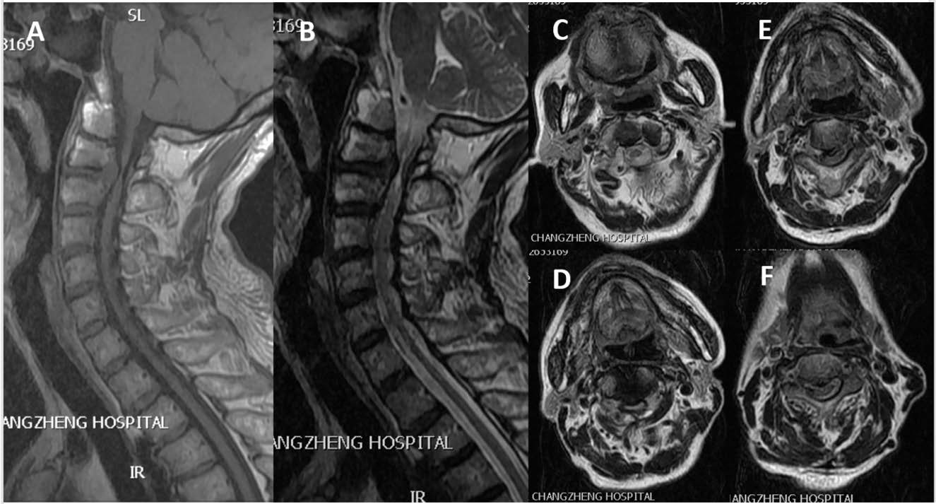
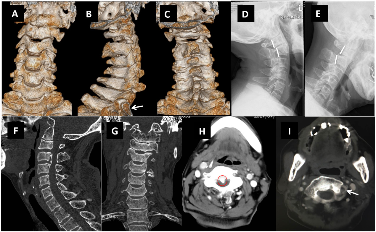
Preoperative three-dimensional reconstruction computed tomography (CT) of the cervical spine indicated hypolordosis with only 6 cervical vertebrae. C1 (atlas) was fused to the occipital bone; C2 was abnormally shaped, lacking the odontoid process, with a posterior arch resembling that of the atlas; C3 displayed normal lateral mass and lamina morphology, with a posterior arch resembling the shape of the axis. The magnetic resonance imaging (MRI) showed the herniated C2/3 intervertebral disc, causing secondary spinal stenosis. The posterior margins of C2 and C3 were significantly misaligned during motion, with a horizontal displacement of 7.7 mm and angular displacement of 19.6°, resulting in C2/3 cervical instability and severe spinal cord compression. OPLL was visible at the C3-C4 level, with the spinal cord showed string-like shape. Diagnosis: (1) cervical spondylotic myelopathy with incomplete paralysis; (2) craniocervical malformation caused by atlantooccipital fusion and absence of odontoid process; and (3) cervical OPLL (Figures 1, 2).

3.1 Anesthesia and position
3.2 Exposure
3.3 Discectomies
3.4 Anterior osteotomy
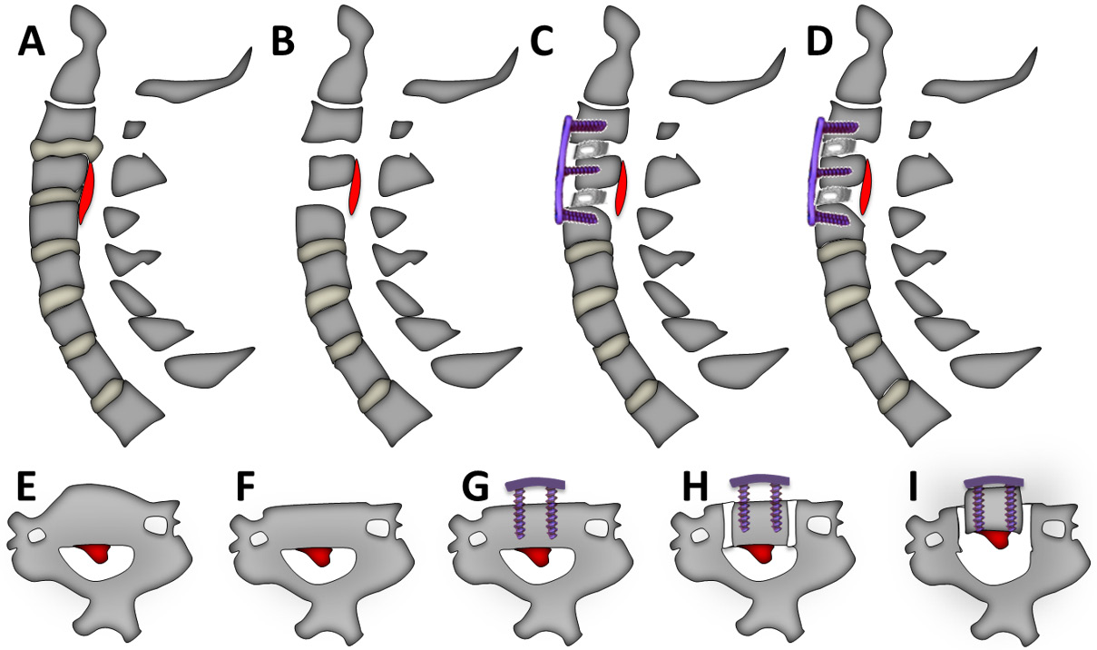
About 3 mm of bone was removed from the anterior side of C3 vertebra using a bone rongeur and an ultrasonic bone curette. Bone wax was applied for hemostasis. A residual vertebral thickness of at least 12 mm was preserved to ensure secure screw fixation during antedisplacement (Figure 3B, 3E, 3F).
3.5 Creation of bilateral grooves
The optimum groove width was determined based on the width of the ossification and the interlaminar distance at each segment from the preoperative 3D reconstruction. The maximum width of the groove was limited to not exceed that of the transverse foramen. Intraoperatively, the actual width was 21.7 mm. Ultrasonic bone curette was used to create a vertical groove along the left side of C3 to a depth reaching the posterior vertebral margin. The bony floor and posterior cortex within the groove were gradually removed using a 1 mm bayonet rongeur and ultrasonic bone curette to expose the posterior longitudinal ligament. A pre-bent titanium plate was fixed onto the anterior surface of C2-C4 with screws. Intraoperative radiography confirmed adequate removal of anterior vertebral bone and correct plate positioning. The right-sided groove of C3 was created in the same manner (Figure 3G, 3H).
3.6 Antedisplacement of vertebrae-ossification complex
Tightening of the C3 screws gradually displaced the C3 vertebra anteriorly into the titanium plate, increasing the spinal canal volume as confirmed by intraoperative radiography. After all screws were fully secured, the surgical field was flushed repeatedly with saline and carefully inspected for hemostasis. A small incision was made adjacent to the groove for placement of a negative-pressure drainage tube. The wound was closed in layers from the aponeurosis to the platysma and subcutaneous tissue, and the skin was finally sealed with absorbable sutures. After confirming patency of the negative pressure drainage, the wound was covered with a sterile dressing, marking the completion of the procedure (Figure 3D, 3I).
The operation lasted 2.3 hours, with an estimated intraoperative blood loss of about 50 ml. There were no neurological deterioration, postoperative hematoma, or other complications, and the surgery was completed successfully. Postoperatively, numbness in the trunk and hands, as well as limb weakness were significantly alleviated. The patient reported no discomfort such as headache, dizziness, nausea, cough, or dyspnea. Postoperative imaging indicated a reduction in T1 slope (41.4° to 25°), an increase in C2-7 sagittal vertical axis (SVA) (4.6 mm to 22.1 mm), and a decrease in C2-7 Cobb angle (40° to 35°). Neurological examination revealed grade 3+ muscle strength in upper limbs and grade 5- in lower limbs. Japanese Orthopaedic Association (JOA) score increased from 6 (before operation) to 10 (after operation) postoperatively, with a neurological recovery rate of 25.3%.
Imaging revealed that the patient had only 6 cervical vertebrae. The lower cervical spine exhibited normal morphology, whereas the upper cervical spine demonstrated structural abnormalities. In a normal cervical spine, the vertebral artery originates from the subclavian artery, ascends along the medial edge of the anterior scalene muscle, enters transverse foramen at C6, and proceeds superiorly to C1. However, MRI of this patient showed that the vertebral artery entered the transverse foramen at 2 segments above T1, corresponding to the fifth segment in this patient’s sixth cervical vertebrae, indicating that this vertebra represented the anatomical C6. Additionally, a 16 mm bony structure was visible at the front of the foramen magnum, bridging the occipital condyles on both sides. These observations suggest that the loss of cervical vertebra in our patient was caused by atlantooccipital fusion. CT images further showed that the posterior arch of C2 and C3 resembled the typical curvature of the atlas and axis, respectively. Notably, the odontoid process was absent at C2, and a pseudojoint was present between C1 and C2. Taken together, these features indicate a rare case of craniocervical malformation with atlantooccipital fusion and absence of the odontoid process.
5.1 Cervical instability
In a normal spine, each pair of adjacent vertebrae and their intervertebral disc, facet joints, and ligaments, form a functional spine unit (FSU). FSUs maintain spinal stability by producing coordinated physiological responses to external and internal forces. However, abnormal increase in the relative displacement of adjacent vertebrae can result in spinal instability. A biomechanical study by White et al. demonstrated that horizontal displacement >3.5 mm or relative rotation >11° between adjacent vertebra can serve as diagnostic criteria for cervical instability [1].
Physiological neck motions require the coordinated movement of all cervical vertebrae. When certain segments lose mobility due to congenital or acquired cervical fusion, motion-induced stress is redistributed to the remaining mobile vertebrae, resulting in increased compensatory movement, mechanical stress, and degenerative changes in the adjacent segments. A biomechanical study of cervical deformity by Eck et al. showed that a reduced number of mobile vertebrae after cervical fusion significantly increases compensatory motion and accelerates degeneration of adjacent segments [2]. Similarly, Pizzutillo et al. reported that fused segments in patients with Klippel-Feil syndrome can lead to kinematic changes and heightened degeneration of adjacent segments [3]. Furthermore, Koyanagi, et al. observed that in patients with continuous OPLL, CDH, cervical instability and injury often occur adjacent to vertebrae immobilized by OPLL [4].
For patients with atlantooccipital fusion, the loss of normal joint structure and function result in the loss of joint movement, often increasing compensatory activity of the atlantoaxial joint and predisposing to atlantoaxial instability [5]. In our case, the patient also had an absence of odontoid process, which further impaired normal atlantoaxial joint functions, while the presence of a pseudojoint at C1/2 not only reduced motion at this level but also increased the mechanical load and compensatory activity in the lower cervical spine. Furthermore, the presence of OPLL at C3 and C4 “fused and fixed” these vertebrae, resulting in reduced C3/4 mobility. We speculated that the increased compensatory activity at C2/3 was an important contributor to the degenerative changes and instability of C2/3. Moreover, persistent cervical instability was also a major cause of exacerbated OPLL [6].
5.2 Posterior approach
In occipitocervical malformations, such as odontoid process malformation with atlantooccipital fusion and skull base depression, spinal compression is typically caused by posterior bony structures. Occipitocervical fusion combined with C1 posterior arch resection is a common surgical treatment for such cases, providing effective decompression and restoration of cervical stability with satisfactory long-term outcomes [7]. In this patient, although the craniocervical malformation was caused by atlantooccipital fusion and absence of the odontoid process, the spinal cord compression was primarily anterior, caused by the herniated C2/3 disc and C3-C4 OPLL. Therefore, treatment for this patient should focus on expanding the cervical canal and relieving spinal compression caused by CDH and OPLL, as well as reestablishing cervical stability and preserving mobility in unaffected vertebrae as much as possible.
Posterior expansive laminoplasty is currently the primary surgical approach with clinically proven efficacy for the treatment of cervical OPLL. By utilizing the physiologic lordosis of the cervical spine and the “bowstring effect” of the spinal cord, this approach can achieve indirect decompression through dorsal shifting of the spinal cord. It also, expansive laminoplasty alone can also largely retain the posterior structures of the cervical spine and its motor functions [8]. However, long-term follow-up of cases with preoperative cervical instability has shown that the lack of immediate stabilization after expansive laminoplasty can result in postoperative kyphosis, OPLL progression, and reclosure of laminar doors, potentially causing recurrent neurological symptoms [9]. Persistent postoperative cervical instability can further exacerbate these symptoms [10]. Previous studies have reported that in 50%–70% of patients undergoing posterior expansive laminoplasty, ossification continues to thicken and elongate at an average rate of 0.3 mm/year and 1 mm/year, respectively. Over 10% of these patients experience recurrence or exacerbation of neurological symptoms [6].
Internal fixation using a screw/rod system can provide superior short-term outcomes for OPLL patients with cervical instability, as it effectively restores cervical lordosis, increases internal fixation strength, and provides immediate postoperative stability. In particular, the posterior pedicle screw fixation achieves strong three-column stability by inserting pedicle screws from the posterior approach into the anterior column of the cervical spine. The perpendicular orientation of paired pedicle screws restricts motion in all planes, thereby restoring stability in patients with cervical instability [11]. However, the posterior approach requires the patient to be in a prone position with the neck flexed, and lateral mass or pedicle screw fixation alone provides only limited correction of cervical lordosis [12]. Moreover, in this patient, the posterior decompression alone could not relieve the anterior compression from the herniated C2/3 disc, leaving residual cord compression and compromising surgical efficacy.
Although the posterior approach is technically straightforward and relatively safe, it is associated with common complications, including C5 nerve root palsy and axial neck/shoulder pain. The incidence of C5 nerve root palsy in cervical OPLL surgery averages 8.3% (3.2%–28.6%), although its pathogenesis is largely unclear [13]. Proposed causes include nerve root stretching dur to dorsal spine cord shift and adhesion between the nerve root and ossification [14, 15]. Axial pain is another common complication, with debated etiology. Hosono et al. believed that it is caused by secondary compression between adjacent laminae and the bulging dura mater following posterior shift-mediated spinal decompression, and that expansion of the decompression range may reduce this risk [16].
In patients with cervical instability, achieving sufficient decompression often requires posterior laminectomy up to C2 and removal of muscles attached to the C2 spinous process. However, postoperative absence of muscle attachment on C2 can impair the maintenance of cervical lordosis, increasing the risk of postoperative kyphosis and OPLL progression. As a result, the presence of preoperative cervical instability, these factors may negatively impact the long-term efficacy of this surgical approach [17].
5.3 Anterior approach
Anterior cervical corpectomy and fusion (ACCF) is an effective surgical approach for directly alleviating spinal compression in OPLL patients. This procedure not only achieves complete decompression via direct resection of the ossified ligament but also restores cervical lordosis by increasing intervertebral height during anterior distraction. ACCF also removes herniated disc tissues during vertebral and ossification resection, making it particularly suitable for OPLL patients with CDH. ACCF provides a good biomechanical environment for the recovery of neurological functions, and hence its neurological improvement rate is superior to that of the posterior approach [18].
However, ACCF is technically more challenging and carries higher surgical risk than the posterior approach. It is generally not recommended for patients with severe involving more than three vertebrae, occupying >50% of the spinal canal, or with ossification thickness >5 mm [19]. Although improvements in surgical techniques and equipment have made anterior approach a possible option for severe OPLL patients, multilevel vertebral resection increases surgical trauma, reconstruction difficulty, and the risks of graft displacement, nonunion, and vertebral collapse [20]. Biomechanical studies have shown that the vertebral and intervertebral discs bear most of the axial load, while approximately 30% is born by small posterior joints. Therefore, corpectomy by ACCF inevitably reduces vertebral stability [21]. In particular, ACCF involving more than two vertebrae substantially increases the risks of steel plate and screw loosening and pseudarthrosis formation. A 31-month follow-up of post-ACCF patients revealed that the failure rate of internal fixation was 6% for two-segment fixation but rose sharply to 71% for three-segment fixation [22]. Our patient had C2/3 instability, CDH, and C3-C4 OPLL. If ACCF was performed with C3 resection, the preoperative instability would likely lead to postoperative stress concentration, pseudarthrosis formation, and fixation failure. More importantly, preoperative instability is a recognized risk factor for accelerated post-ACCF degeneration of adjacent segments [23].
Despite improvements in surgical techniques and instrumentation, anterior approaches such as ACCF remain high-risk procedures with a relatively high complication rate. Cerebrospinal fluid (CSF) leakage is a common surgical complication of ACCF, most often resulting from dural mater tearing during separation of the dura mater from an adherent posterior longitudinal ligament [24]. In severe OPLL, dural ossification may be present, and resection of the ossified mass via ACCF can cause substantial dural injury, leading to sustained postoperative CSF leakage and impaired recovery [25]. Our patient had OPLL and upper cervical malformation with an ossification thickness of 4.6 mm and a sagittal canal diameter of 3 mm at the most affected segments. Since the anterior approach is more technically challenging, the risk of intraoperative dural or spinal cord injury—potentially resulting in CSF leak or even paraplegia—would be markedly elevated.
5.4 Combined anterior and posterior approach
Based on the conditions of our patient, a combined anterior-posterior approach could theoretically provide ideal spinal decompression. Specifically, posterior expansive laminoplasty can first be performed to expand the volume of the cervical spinal canal, allowing greater dorsal shift of the spinal cord and indirect reduction of anterior compression from ossification. As the spinal cord shifts towards the back, the increased anterior space of the cervical spine provides more operating space for anterior corpectomy and reduces the risk of intraoperative complications. The anterior approach can then be performed to completely remove residual compression caused by OPLL and CDH, while anterior bone graft fusion restores cervical stability and prevents instability resulting from posterior column compromise [26]. The major advantage of this combined approach is the ability to achieve complete decompression while minimizing the risk of surgical complication [27]. However, this procedure is lengthy, highly invasive, and associated with substantial intraoperative blood loss. Prolonged manipulation of the spinal cord may also introduce additional neurological risks. In addition, this approach requires good patient tolerance, and in some cases, staged surgery may be necessary [28].
5.5 ACAF
ACAF is a surgical technique that directly expands the spinal canal by anteriorly displacing the vertebra-ossification-dura mater complex. This forward shift restores the spinal canal volume, relieves compression on the spinal cord and nerve roots, and re-establish the normal vertebral structure and natural physiological environment for nerve tissues. ACAF combines the direct, complete decompression and safety features of the traditional anterior and posterior approaches.
In terms of postoperative stability, ACAF involves bilateral isolation and en bloc anterior displacement of the OPLL complex following anterior cervical disectomy and fusion (ACDF). Although this inevitably compromises the stability of the anterior and middle cervical columns, biomechanical studies have indicated that immediate postoperative stability with ACAF is superior to ACCF, even though both employ anterior plate and screw fixation [29]. This advantage is especially apparent in cases with multilevel segments and may be attributable to the fact that ACAF achieves decompression without resection of OPLL complex, thereby preserving more structural integrity.
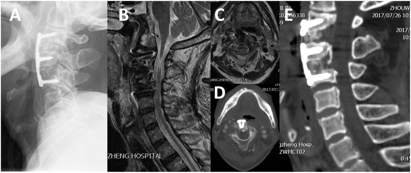
For OPLL patients with cervical instability, ACAF is better than ACCF in preventing instability exacerbation and reestablishing cervical stability. In the present case, plate fixation at C2-4 partially restricted cervical spine activity but effectively eliminated instability-induced degeneration. Postoperative imaging confirmed secure internal fixation and complete resolution of cervical instability (Figure 4).
In terms of decompression efficacy, en bloc isolation and anterior displacement of the involved segments following ACDF can directly relieve compression caused by CDH, allowing ACAF to achieve direct and complete spinal decompression. In this case, he procedure effectively resolved compression from C2/3 CDH. In addition, the width of bilateral osteotomies and intervertebral space for decompression in ACAF can be up to 18–22 mm - wider than the spinal cord and the conventional 14–16 mm in ACCF - and can extend to the medial margins of the nerve root exits. This allows simultaneous decompression of the spinal cord and nerve roots. Imaging examination of our patient showed a sagittal canal diameter of 14.9 mm at the C3, indicating complete spinal decompression.
Given that ACAF is a direct anterior decompression approach that does not require dorsal shift of the spinal cord, it effectively avoids postoperative C5 nerve root palsy and axial neck/shoulder pain, which are common in the posterior approach. The advantage of ACAF over traditional anterior approaches is that it does not require separation of the posterior longitudinal ligament from the dura mater; instead, the entire ossification complex is directly lifted anteriorly through the “safe grooves” created by bilateral osteotomies. In the event of a CSF leakage, these grooves can be sealed with autologous fascial tissues or artificial dura mater. This procedure is simple and effective as the entire vertebra-ossification complex is handled en bloc [30]. At 3-month follow-up, the patient demonstrated satisfactory recovery of neurologic functions without the complications such as nerve root palsy and axial pain.
Despite ACAF being a direct anterior decompression procedure, its operative field is close to the Luschka’s joints lateral to the spinal cord, and the absence of direct ossification resection reduces the risk of intraoperative spinal cord injury. Osteotomies during ACAF are performed using an ultrasonic bone curette or high-speed drill, both of which provide certain hemostatic effects during bone cutting. Bone wax can also be applied if increased bone bleeding is observed. In the present case, ultrasonic bone curette-assisted osteotomy was used to make the grooves, with a total operative time of 2.3 hours, estimated blood loss of about 50 ml, and no postoperative hematoma or worsening of neurologic functions.
ACAF is an emerging surgical technique that integrates the direct, complete decompression from traditional anterior approaches with the safety profile of posterior approaches. Although further clinical studies are needed to validate its long-term efficacy and safety, its favorable biomechanical properties, complete decompression, and low risk of surgical complications make ACAF a promising treatment for OPLL with cervical instability.
Cervical instability may play a pivotal role in the pathogenesis of spinal degeneration and related symptoms. In this case, the herniated C2/3 disc and C3-C4 OPLL were the direct cause of nerve compression, likely secondary to biomechanical alterations from atlanto-occipital fusion and atlantoaxial instability, with stress transmission along the cervical spine [31]. Cervical spine instability is also recognized as an important cause of ligament ossification, and thus, “only-fixation” strategies without decompression have been reported to yeild good results in selected OPLL patients [32, 33]. In this case, ACAF achieved direct anterior decompression without removing the ossification, while restoring the stability at the compressed segments and eliminating a major driver of ossification progression. Compared with traditional ACCF and laminoplasty, ACAF offers a superior balance between decompression efficacy and surgical safety.
Anterior controllable antedisplacement and fusion (ACAF) represents an innovative surgical approach that combines the direct decompression efficacy of anterior approach with the safety advantages of posterior indirect decompression. It offers favorable biomechanical attributes, the potential for complete decompression, and a relatively low incidence of surgical complications. As such, ACAF may serve as a valuable treatment alternative for patients with ossification of the posterior longitudinal ligament (OPLL) accompanied by cervical instability. Nevertheless, additional research and clinical experience are needed to fully establish its long - term efficacy and optimal applications.
Author contributions
Xi Luo (First Author): Conceived and designed the study, conducted the experiments, analyzed the data, and drafted the initial manuscript. Jingchuan Sun (Corresponding Author): Supervised the study, provided critical insights and guidance throughout the research process, and reviewed and revised the manuscript.
Funding
No funding was received for the conduct of this study or the writing of this manuscript.
Data availability
The datasets generated and analyzed during the current study are available from the corresponding author on reasonable request.
Ethics approval and consent to participate
This study was approved by the Ethics Committee of the corresponding hospital (Shanghai Changzheng Hospital, 2017SL040). All procedures performed in this study were in accordance with the ethical standards of the institutional research committee and with the 1964 Helsinki declaration and its later amendments. Written informed consent was obtained from all individual participants included in the study.
Consent for publication
All authors have read and approved the final version of the manuscript, and consented to the publication of this research work.
Competing interests
The authors declare that there are no competing financial or non-financial interests in relation to the work described in this manuscript.
Acknowledgements
Not applicable.
[1] White A, Southwick W, Panjabi M. Clinical instability in the lower cervical spine a review of past and current concepts. Spine. 1976;1(1):15-27. https://doi.org/10.1097/00007632-197603000-00003
[2] Eck JC, Humphreys SC, Lim TH, Jeong ST, Kim JG, Hodges SD, et al. Biomechanical study on the effect of cervical spine fusion on adjacent-level intradiscal pressure and segmental motion. Spine (Phila Pa 1976). 2002 Nov 15;27(22):2431-2434. https://doi.org/10.1097/00007632-200211150-00003
[3] Pizzutillo PD, Woods M, Nicholson L, MacEwen GD. Risk factors in Klippel-Feil syndrome. Spine (Phila Pa 1976). 1994 Sep 15;19(18):2110-2116. https://doi.org/10.1097/00007632-199409150-00020
[4] Koyanagi I, Iwasaki Y, Hida K, Imamura H, Fujimoto S, Akino M. Acute cervical cord injury associated with ossification of the posterior longitudinal ligament. Neurosurgery. 2003 Oct;53(4):887-891; discussion 891-892. https://doi.org/10.1227/01.neu.0000083590.84053.cc
[5] Lee DH, Cho ST, Kang HW, Park S, Hwang CJ, Cho JH. Comparison between atlantoaxial and occipitocervical fusion: Clinical implications of restoring the atlanto-occipital joint. Spine J. 2025 Apr;25(4):749-755. https://doi.org/10.1016/j.spinee.2024.11.002
[6] Ogawa Y, Toyama Y, Chiba K, Matsumoto M, Nakamura M, Takaishi H, et al. Long-term results of expansive open-door laminoplasty for ossification of the posterior longitudinal ligament of the cervical spine. J Neurosurg Spine. 2004 Sep;1(2):168-174. https://doi.org/10.3171/spi.2004.1.2.0168
[7] Hankinson TC, Grunstein E, Gardner P, Spinks TJ, Anderson RC. Transnasal odontoid resection followed by posterior decompression and occipitocervical fusion in children with Chiari malformation Type I and ventral brainstem compression. J Neurosurg Pediatr. 2010 Jun;5(6):549-553. https://doi.org/10.3171/2010.2.PEDS09362
[8] Liu X, Yuan S, Tian Y, Zheng Y, Li J. Expansive open‐door laminoplasty and selective anterior cervical decompression and fusion for treatment of multilevel cervical spondylotic myelopathy. Orthop Surg. 2011 Aug;3(3):161-166. https://doi.org/10.1111/j.1757-7861.2011.00143.x
[9] Chiba K, Ogawa Y, Ishii K, Takaishi H, Nakamura M, Maruiwa H, et al. Long-term results of expansive open-door laminoplasty for cervical myelopathy--average 14-year follow-up study. Spine (Phila Pa 1976). 2006 Dec 15;31(26):2998-3005. https://doi.org/10.1097/01.brs.0000250307.78987.6b
[10] Hori T, Kawaguchi Y, Kimura T. How does the ossification area of the posterior longitudinal ligament thicken following cervical laminoplasty? Spine (Phila Pa 1976). 2007 Sep 1;32(19):E551-E556. https://doi.org/10.1097/BRS.0b013e31814614f3
[11] Kothe R, Rüther W, Schneider E, Linke B. Biomechanical analysis of transpedicular screw fixation in the subaxial cervical spine. Spine (Phila Pa 1976). 2004 Sep 1;29(17):1869-1875. https://doi.org/10.1097/01.brs.0000137287.67388.0b
[12] Kim SH, Kim JH, Kwon JW, Kim HS, Moon SH, Suk KS, et al. Assessment of biomechanical advantages in combined anterior–posterior cervical spine surgery by radiological outcomes: Pedicle screws over lateral mass screws. J Clin Med. 2023 Apr 29;12(9):3201. https://doi.org/10.3390/jcm12093201
[13] Sakaura H, Hosono N, Mukai Y, Ishii T, Yoshikawa H. C5 palsy after decompression surgery for cervical myelopathy: Review of the literature. Spine (Phila Pa 1976). 2003 Nov 1;28(21):2447-2451. https://doi.org/10.1097/01.BRS.0000090833.96168.3F
[14] Bak AB, Moghaddamjou A, Alvi M, Ahn H, Farhadi HF, Shaffrey CI, et al. Postoperative C5 palsy after anterior or posterior decompression for degenerative cervical myelopathy: A subgroup analysis of the multicenter, prospective, randomized, Phase III, CSM-Protect clinical trial. Spine (Phila Pa 1976). 2024 Oct 15;49(20):1410-1416. https://doi.org/10.1097/BRS.0000000000005007
[15] Chen Y, Chen D, Wang X, Guo Y, He Z. C5 palsy after laminectomy and posterior cervical fixation for ossification of posterior longitudinal ligament. J Spinal Disord Tech. 2007 Oct;20(7):533-535. https://doi.org/10.1097/BSD.0b013e318042b655
[16] Hosono N, Sakaura H, Mukai Y, Fujii R, Yoshikawa H. C3-6 laminoplasty takes over C3-7 laminoplasty with significantly lower incidence of axial neck pain. Eur Spine J. 2006 Sep;15(9):1375-1379. https://doi.org/10.1007/s00586-006-0089-9
[17] An S, Lee JB, Lee S, Oh Y, Park JY, Hur JW. True continuous segment of ossification of posterior longitudinal ligament is protective against postoperative early kyphosis progression after laminoplasty. Neurosurgery. 2023 Nov 22;94(5):933-943. https://doi.org/10.1227/neu.0000000000002773
[18] Monk SH, O’Brien M, Perle S, Bohl M, Finger F, Chewning SJ, et al. Ten-year experience of skip anterior cervical corpectomy and fusion. Int J Spine Surg. 2023 Apr;17(2):258-264. https://doi.org/10.14444/8417
[19] Wang S, Xiang Y, Wang X, Li H, Hou Y, Zhao H, et al. Anterior corpectomy comparing to posterior decompression surgery for the treatment of multi-level ossification of posterior longitudinal ligament: A meta-analysis. Int J Surg. 2017 Apr;40:91-96. https://doi.org/10.1016/j.ijsu.2017.02.058
[20] Lee Y, Trenchfield DB, Berthiaume E, Tomlak AB, Narayanan R, Brush P, et al. A comparison of clinical outcomes between anterior cervical discectomy and fusion versus posterior cervical laminoplasty for multilevel cervical myelopathy. Clin Spine Surg. 2024 Dec 1;37(10):E529-E535. https://doi.org/10.1097/BSD.0000000000001634
[21] Lofrese G, Trungu S, Scerrati A, De Bonis P, Cultrera F, Mongardi L, et al. Two-level corpectomy and fusion vs. three-level anterior cervical discectomy and fusion without plating: Long-term clinical and radiological outcomes in a multicentric retrospective analysis. Life (Basel). 2023 Jul 14;13(7):1564. https://doi.org/10.3390/life13071564
[22] Sasso RC, Ruggiero RA, Reilly TM, Hall PV. Early reconstruction failures after multilevel cervical corpectomy. Spine (Phila Pa 1976). 2003 Jan 15;28(2):140-142. https://doi.org/10.1097/00007632-200301150-00009
[23] Chung JY, Kim SK, Jung ST, Lee KB. Clinical adjacent-segment pathology after anterior cervical discectomy and fusion: Results after a minimum of 10-year follow-up. Spine J. 2014 Oct 1;14(10):2290-2298. https://doi.org/10.1016/j.spinee.2014.01.027
[24] Wang M, Yang G, Zhou B, Cao Z, Li Y, Tan J, et al. Anterior cervical controllable antedisplacement and fusion (ACAF) versus anterior cervical corpectomy and fusion (ACCF) for ossification of the cervical posterior longitudinal ligament (OPLL) in Chinese population: A systematic review and meta-analysis. Neurosurg Rev. 2024 Oct 11;47(1):783. https://doi.org/10.1007/s10143-024-02977-x
[25] Mazur M, Jost GF, Schmidt MH, Bisson EF. Management of cerebrospinal fluid leaks after anterior decompression for ossification of the posterior longitudinal ligament: A review of the literature. Neurosurg Focus. 2011 Mar;30(3):E13. https://doi.org/10.3171/2010.12.FOCUS10255
[26] Matsumoto M, Chiba K, Toyama Y, Takeshita K, Seichi A, Nakamura K, et al. Surgical results and related factors for ossification of posterior longitudinal ligament of the thoracic spine: A multi-institutional retrospective study. Spine (Phila Pa 1976). 2008 Apr 20;33(9):1034-1041. https://doi.org/10.1097/BRS.0b013e31816c913b
[27] Chen H, Chen D, Zhou S, Sang L, Wu J, Huang F. Combined anterior and posterior approach in treatment of ankylosing spondylitis-associated cervical fractures: A systematic review and meta-analysis. Eur Spine J. 2023 Jan;32(1):27-37. https://doi.org/10.1007/s00586-022-07435-0
[28] Kawaguchi Y, Nakano M, Yasuda T, Seki S, Hori T, Kimura T. Anterior decompressive surgery after cervical laminoplasty in patients with ossification of the posterior longitudinal ligament. Spine J. 2014 Jun 1;14(6):955-963. https://doi.org/10.1016/j.spinee.2013.07.457
[29] Kong Q, Li F, Yan C, Sun J, Sun P, Ou-Yang J, et al. Biomechanical comparison of anterior cervical corpectomy decompression and fusion, anterior cervical discectomy and fusion, and anterior controllable antedisplacement and fusion in the surgical treatment of multilevel cervical spondylotic myelopathy: A finite element analysis. Orthop Surg. 2024 Mar;16(3):687-699. https://doi.org/10.1111/os.13994
[30] Sun J, Sun K, Wang Y, Shi J, Yang H, Guo Y, et al. Quantitative anterior enlargement of the spinal canal by anterior controllable antedisplacement and fusion for the treatment of cervical ossification of the posterior longitudinal ligament with myelopathy. World Neurosurg. 2018 Dec;120:e1098-e1106. https://doi.org/10.1016/j.wneu.2018.08.233
[31] Vij N, Tolson H, Kiernan H, Agusala V, Viswanath O, Urits I. Pathoanatomy, biomechanics, and treatment of upper cervical ligamentous instability: A literature review. Orthop Rev (Pavia). 2022 Aug 5;14(3):37099. https://doi.org/10.52965/001c.37099
[32] Luo X, Sun K, Zhu J, Wang S, Wang Y, Sun J, et al. Analysis of intervertebral disc degeneration in patients with ossification of the posterior longitudinal ligament. Quant Imaging Med Surg. 2022 Mar;12(3):1919-1928. https://doi.org/10.21037/qims-21-154
[33] Goel A, Vutha R, Shah A, Prasad A, Shukla AK, Maheshwari S. Defining role of atlantoaxial and subaxial spinal instability in the pathogenesis of cervical spinal degeneration: Experience with “only-fixation” without any decompression as treatment in 374 cases over 10 years. J Craniovertebr Junction Spine. 2024 Jan-Mar;15(1):74-82. https://doi.org/10.4103/jcvjs.jcvjs_11_24


 On This Page
 CITE
CITE